Copyright
©2011 Baishideng Publishing Group Co.
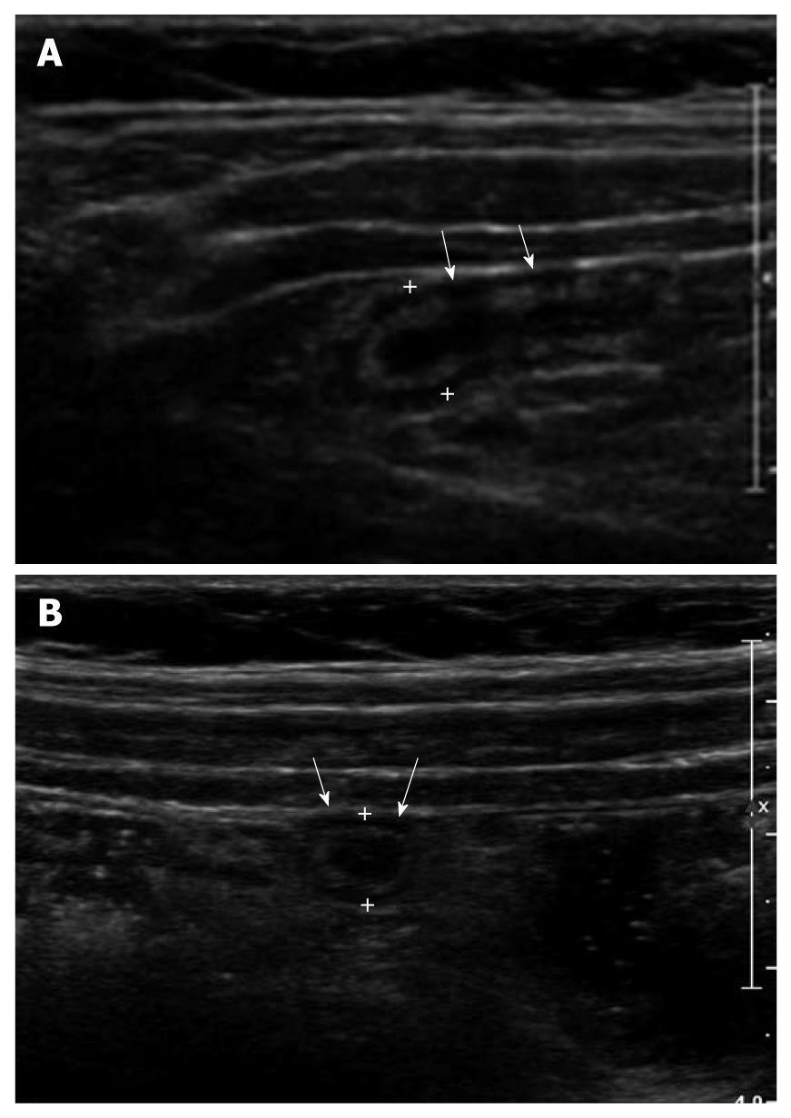
Figure 6 A 10-year-old male with a non-obstructive acute appendicitis.
A: Thickening of appendiceal wall (arrows) with a maximal outer diameter (MOD) of 3 mm and some irregularity of the submucosal echogenic layer. However, no evidence of luminal distention and intraluminal appendicolith was found; B: MOD of inflamed appendix (arrows) was measured as 7.1 mm.
- Citation: Park NH, Oh HE, Park HJ, Park JY. Ultrasonography of normal and abnormal appendix in children. World J Radiol 2011; 3(4): 85-91
- URL: https://www.wjgnet.com/1949-8470/full/v3/i4/85.htm
- DOI: https://dx.doi.org/10.4329/wjr.v3.i4.85


