Copyright
©2011 Baishideng Publishing Group Co.
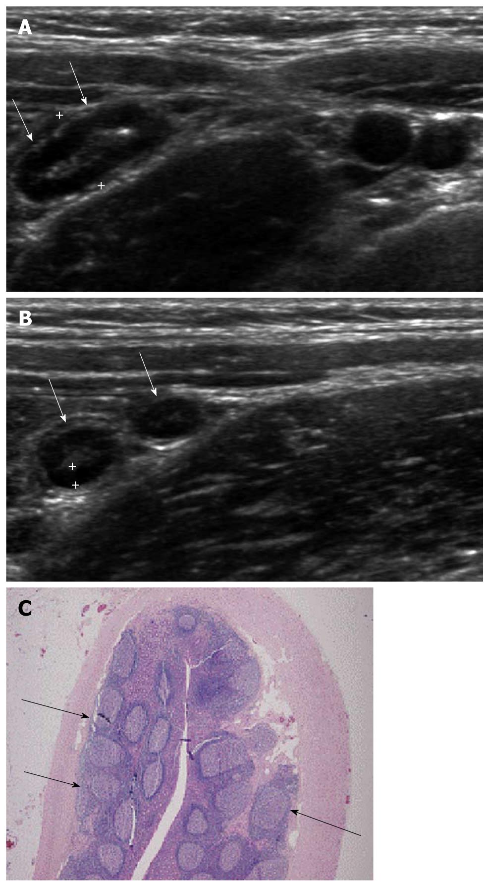
Figure 2 Ultrasonographic and histologic findings of mucosal lymphoid hyperplasia of the appendix.
A: Normal appendix with thick inner hypoechoic band was seen (arrows). The maximal outer diameter was measured as 7.3 mm; B: The thickness of the inner hypoechoic band (cursors) of the appendix (arrows) was measured as 1.4 mm; C: Prominent lymphoid follicles (arrows) were found (× 20, H-E stain).
- Citation: Park NH, Oh HE, Park HJ, Park JY. Ultrasonography of normal and abnormal appendix in children. World J Radiol 2011; 3(4): 85-91
- URL: https://www.wjgnet.com/1949-8470/full/v3/i4/85.htm
- DOI: https://dx.doi.org/10.4329/wjr.v3.i4.85


