Copyright
©2011 Baishideng Publishing Group Co.
World J Radiol. Dec 28, 2011; 3(12): 298-305
Published online Dec 28, 2011. doi: 10.4329/wjr.v3.i12.298
Published online Dec 28, 2011. doi: 10.4329/wjr.v3.i12.298
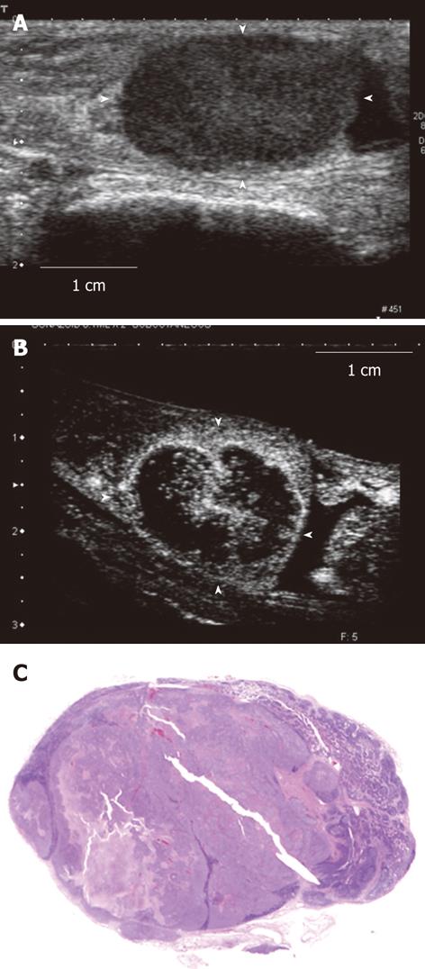
Figure 3 Contrast-enhanced ultrasonography image and histopathological image of the tumor-induced lymph node enlargement model.
This is the tumor-induced lymph node enlargement model at 28 d after VX2 tumor was implanted (Model 1). A: The enlarged popliteal lymph node with a diameter of 18 mm that was seen in the B mode ultrasound image. This lymph node shown hypoechoic mass; B: Image of the popliteal lymph node that was imaged after the contrast agent was administered in the periphery of the primary tumor lesion. The central area is large and defective and so only the periphery of the lymph node was imaged; C: Histopathological image (hematoxylin-eosin stain) of the lymph node that was extracted. A large metastatic tumor lesion was seen in the center.
- Citation: Aoki T, Moriyasu F, Yamamoto K, Shimizu M, Yamada M, Imai Y. Image of tumor metastasis and inflammatory lymph node enlargement by contrast-enhanced ultrasonography. World J Radiol 2011; 3(12): 298-305
- URL: https://www.wjgnet.com/1949-8470/full/v3/i12/298.htm
- DOI: https://dx.doi.org/10.4329/wjr.v3.i12.298


