Copyright
©2011 Baishideng Publishing Group Co.
World J Radiol. Dec 28, 2011; 3(12): 289-297
Published online Dec 28, 2011. doi: 10.4329/wjr.v3.i12.289
Published online Dec 28, 2011. doi: 10.4329/wjr.v3.i12.289
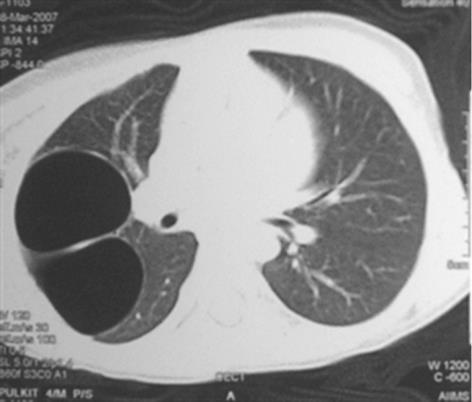
Figure 14 Congenital cystic adenomatoid malformation.
Axial image in the lung window depicts the presence of thin walled and septated air-filled cysts (arrow) in the right upper lobe, causing mass effect on the adjacent lung suggestive of congenital cystic adenomatoid malformation type-1.
- Citation: Sundarakumar DK, Bhalla AS, Sharma R, Gupta AK, Kabra SK, Jagia P. Multidetector computed tomography imaging of congenital anomalies of major airways: A pictorial essay. World J Radiol 2011; 3(12): 289-297
- URL: https://www.wjgnet.com/1949-8470/full/v3/i12/289.htm
- DOI: https://dx.doi.org/10.4329/wjr.v3.i12.289


