Copyright
©2011 Baishideng Publishing Group Co.
World J Radiol. Dec 28, 2011; 3(12): 289-297
Published online Dec 28, 2011. doi: 10.4329/wjr.v3.i12.289
Published online Dec 28, 2011. doi: 10.4329/wjr.v3.i12.289
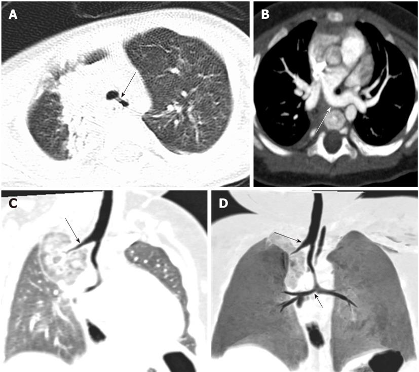
Figure 7 Tracheal bronchus with pulmonary artery sling.
Axial image in the lung window (A) shows the right upper lobe bronchus (long arrow) seen arising directly from the trachea. Axial image in mediastinal (B) shows the left pulmonary artery sling arising from the right branch pulmonary artery (long arrow). There is a separate origin of right upper lobe bronchus (long arrows) from the trachea, and the carinal angle is obtuse as seen in multiplanar reformatted images (C) and minimal intensity projection images (minip) (D) (short arrow). These findings were best depicted by minIP.
- Citation: Sundarakumar DK, Bhalla AS, Sharma R, Gupta AK, Kabra SK, Jagia P. Multidetector computed tomography imaging of congenital anomalies of major airways: A pictorial essay. World J Radiol 2011; 3(12): 289-297
- URL: https://www.wjgnet.com/1949-8470/full/v3/i12/289.htm
- DOI: https://dx.doi.org/10.4329/wjr.v3.i12.289


