Copyright
©2011 Baishideng Publishing Group Co.
World J Radiol. Dec 28, 2011; 3(12): 289-297
Published online Dec 28, 2011. doi: 10.4329/wjr.v3.i12.289
Published online Dec 28, 2011. doi: 10.4329/wjr.v3.i12.289
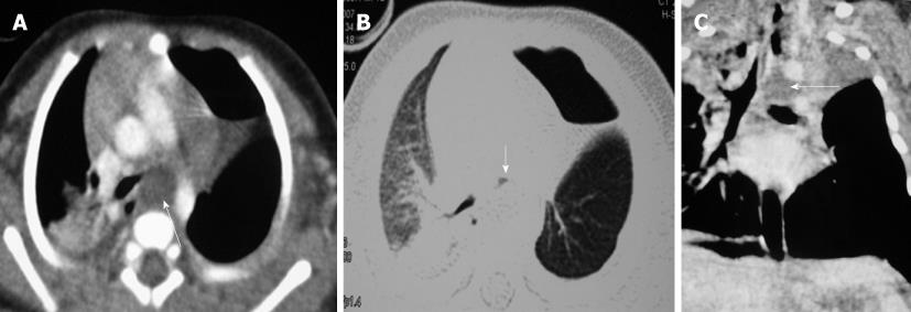
Figure 2 Foregut duplication cyst.
Axial images in the mediastinal (A) and lung (B) window and coronal multiplanar reformatted images (C) showing a fluid-attenuating lesion (long arrows) in the mediastinum compressing the left main bronchus (short arrow) with hyperinflation of left lower lobe. Hydropneumothorax in the left side was due to post-surgical change.
- Citation: Sundarakumar DK, Bhalla AS, Sharma R, Gupta AK, Kabra SK, Jagia P. Multidetector computed tomography imaging of congenital anomalies of major airways: A pictorial essay. World J Radiol 2011; 3(12): 289-297
- URL: https://www.wjgnet.com/1949-8470/full/v3/i12/289.htm
- DOI: https://dx.doi.org/10.4329/wjr.v3.i12.289


