Copyright
©2011 Baishideng Publishing Group Co.
World J Radiol. Dec 28, 2011; 3(12): 279-288
Published online Dec 28, 2011. doi: 10.4329/wjr.v3.i12.279
Published online Dec 28, 2011. doi: 10.4329/wjr.v3.i12.279
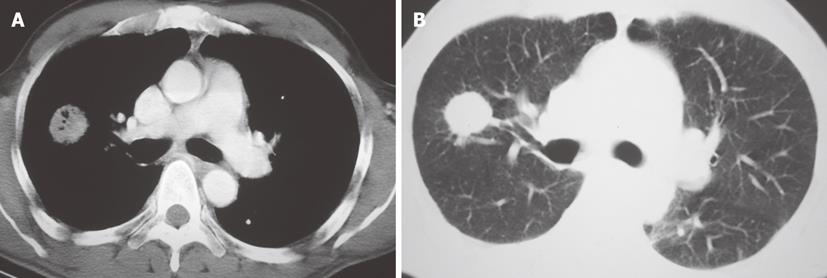
Figure 12 Contrast enhanced chest CT.
A: Mediastinum window, axial image at the level of the pulmonary arteries demonstrates a soft tissue density mass with air-bronchogram in the right lung. B: Lung window image at the same level shows the spiculated contour of the lung mass.
- Citation: Restrepo CS, Chen MM, Martinez-Jimenez S, Carrillo J, Restrepo C. Chest neoplasms with infectious etiologies. World J Radiol 2011; 3(12): 279-288
- URL: https://www.wjgnet.com/1949-8470/full/v3/i12/279.htm
- DOI: https://dx.doi.org/10.4329/wjr.v3.i12.279


