Copyright
©2011 Baishideng Publishing Group Co.
World J Radiol. Dec 28, 2011; 3(12): 279-288
Published online Dec 28, 2011. doi: 10.4329/wjr.v3.i12.279
Published online Dec 28, 2011. doi: 10.4329/wjr.v3.i12.279
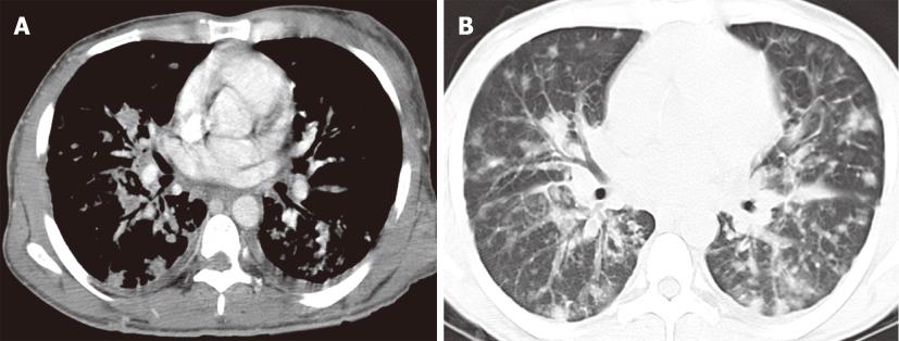
Figure 6 Kaposi sarcoma in a 37-year-old male with acquired immunodeficiency syndrome.
Contrast enhanced computed tomography of the chest, mediastinal window (A) and lung window (B) images demonstrate innumerable peribronchovascular and peripheral pulmonary nodules throughout the bilateral lungs. Enhancing skin lesions and bilateral pleural effusion are also noted.
- Citation: Restrepo CS, Chen MM, Martinez-Jimenez S, Carrillo J, Restrepo C. Chest neoplasms with infectious etiologies. World J Radiol 2011; 3(12): 279-288
- URL: https://www.wjgnet.com/1949-8470/full/v3/i12/279.htm
- DOI: https://dx.doi.org/10.4329/wjr.v3.i12.279


