Copyright
©2011 Baishideng Publishing Group Co.
World J Radiol. Dec 28, 2011; 3(12): 279-288
Published online Dec 28, 2011. doi: 10.4329/wjr.v3.i12.279
Published online Dec 28, 2011. doi: 10.4329/wjr.v3.i12.279
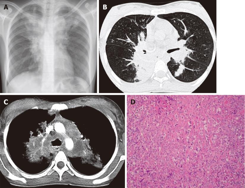
Figure 1 Hodgkin’s lymphoma.
A 23-year-old woman with a four-mo history of dry cough and chest pain. A: Chest X-ray shows mediastinal widening and upper lobe parenchymal opacities; B and C: Contrast-enhanced computed tomography confirms lymphadenopathy involving the mediastinum and infiltrative masses in the bilateral upper lobes; D: Photomicrograph (HE stain). Nodular sclerosing HL lymph node. Fibrous bands divide the lymphoid infiltrate into nodules and contain Hodgkin cells.
- Citation: Restrepo CS, Chen MM, Martinez-Jimenez S, Carrillo J, Restrepo C. Chest neoplasms with infectious etiologies. World J Radiol 2011; 3(12): 279-288
- URL: https://www.wjgnet.com/1949-8470/full/v3/i12/279.htm
- DOI: https://dx.doi.org/10.4329/wjr.v3.i12.279


