Copyright
©2011 Baishideng Publishing Group Co.
World J Radiol. Nov 28, 2011; 3(11): 266-272
Published online Nov 28, 2011. doi: 10.4329/wjr.v3.i11.266
Published online Nov 28, 2011. doi: 10.4329/wjr.v3.i11.266
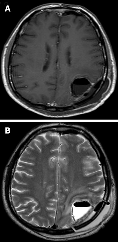
Figure 5 Appearance of Gliadel wafers in the acute phase.
A: Axial post-contrast T1 weighted image (TR, 509 ms; TE, 14 ms) demonstrates non-enhancing T1 hypointense linear structures (arrows) reflecting the Gliadel wafers in the resection cavity site. Post-treatment changes are also seen; B: Axial T2-weighted image (TR, 3000 ms; TE, 80 ms) demonstrates the wafers (arrows) to also be hypointense on T2 and are well seen within the T2 hyperintense fluid-filled resection cavity.
- Citation: Colen RR, Zinn PO, Hazany S, Do-Dai D, Wu JK, Yao K, Zhu JJ. Magnetic resonance imaging appearance and changes on intracavitary Gliadel wafer placement: A pilot study. World J Radiol 2011; 3(11): 266-272
- URL: https://www.wjgnet.com/1949-8470/full/v3/i11/266.htm
- DOI: https://dx.doi.org/10.4329/wjr.v3.i11.266


