Copyright
©2011 Baishideng Publishing Group Co.
World J Radiol. Nov 28, 2011; 3(11): 266-272
Published online Nov 28, 2011. doi: 10.4329/wjr.v3.i11.266
Published online Nov 28, 2011. doi: 10.4329/wjr.v3.i11.266
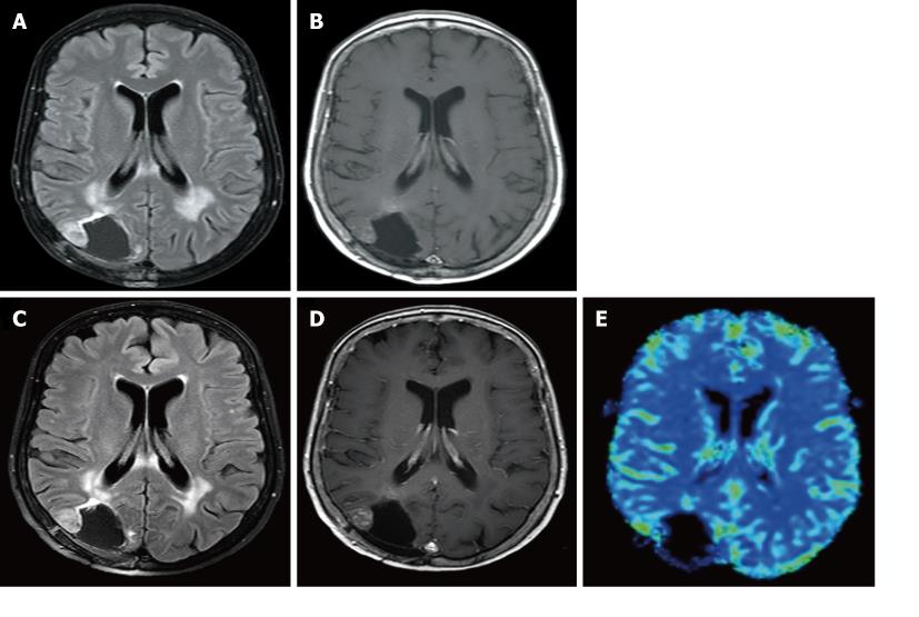
Figure 3 MR appearance in a 59-year-old male patient with tumor progression (Patient 2).
A: One month post-operative, (A) FLAIR (TR, 11,000 ms; TE, 140 ms; TI, 2600 ms) and (B) T1 weighted image (T1WI) (TR,509 ms; TE, 14 ms) demonstrate an increase in FLAIR hyperintensity and enhancement, respectively, surrounding the resection cavity site. Five months post-operative; C, D: (C) FLAIR (TR, 11,000 ms; TE, 140 ms; TI, 2600 ms) and (D) T1WI (TR,509 ms; TE, 14 ms) demonstrate a further increase in FLAIR hyperintensity and enhancement, respectively, surrounding the resection cavity site; E: MR perfusion (rCBV maps) demonstrates increased rCBV in the right posterolateral, anterior and medial aspects (arrows) of the resection cavity site confirming true tumor recurrence rather than post-treatment changes.
- Citation: Colen RR, Zinn PO, Hazany S, Do-Dai D, Wu JK, Yao K, Zhu JJ. Magnetic resonance imaging appearance and changes on intracavitary Gliadel wafer placement: A pilot study. World J Radiol 2011; 3(11): 266-272
- URL: https://www.wjgnet.com/1949-8470/full/v3/i11/266.htm
- DOI: https://dx.doi.org/10.4329/wjr.v3.i11.266


