Copyright
©2011 Baishideng Publishing Group Co.
World J Radiol. Nov 28, 2011; 3(11): 266-272
Published online Nov 28, 2011. doi: 10.4329/wjr.v3.i11.266
Published online Nov 28, 2011. doi: 10.4329/wjr.v3.i11.266
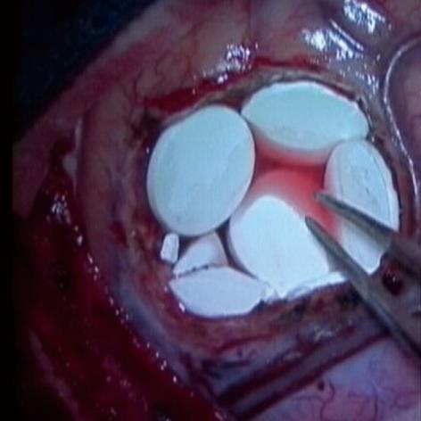
Figure 1 Intracavitary Gliadel wafer placement.
This image, taken at time of Gliadel wafer placement in the operating room, demonstrates the lining of wafers (white round structures) along the wall of the resection cavity site.
- Citation: Colen RR, Zinn PO, Hazany S, Do-Dai D, Wu JK, Yao K, Zhu JJ. Magnetic resonance imaging appearance and changes on intracavitary Gliadel wafer placement: A pilot study. World J Radiol 2011; 3(11): 266-272
- URL: https://www.wjgnet.com/1949-8470/full/v3/i11/266.htm
- DOI: https://dx.doi.org/10.4329/wjr.v3.i11.266


