Copyright
©2011 Baishideng Publishing Group Co.
World J Radiol. Oct 28, 2011; 3(10): 246-248
Published online Oct 28, 2011. doi: 10.4329/wjr.v3.i10.246
Published online Oct 28, 2011. doi: 10.4329/wjr.v3.i10.246
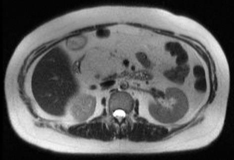
Figure 2 T1 weighted magnetic resonance imaging phase.
This cross sectional image reveals a homogenous fatty containing lesion in the head of pancreas.
- Citation: Lee SY, Thng CH, Chow PK. Lipoma of the pancreas, a case report and a review of the literature. World J Radiol 2011; 3(10): 246-248
- URL: https://www.wjgnet.com/1949-8470/full/v3/i10/246.htm
- DOI: https://dx.doi.org/10.4329/wjr.v3.i10.246


