Copyright
©2010 Baishideng Publishing Group Co.
World J Radiol. Sep 28, 2010; 2(9): 358-367
Published online Sep 28, 2010. doi: 10.4329/wjr.v2.i9.358
Published online Sep 28, 2010. doi: 10.4329/wjr.v2.i9.358
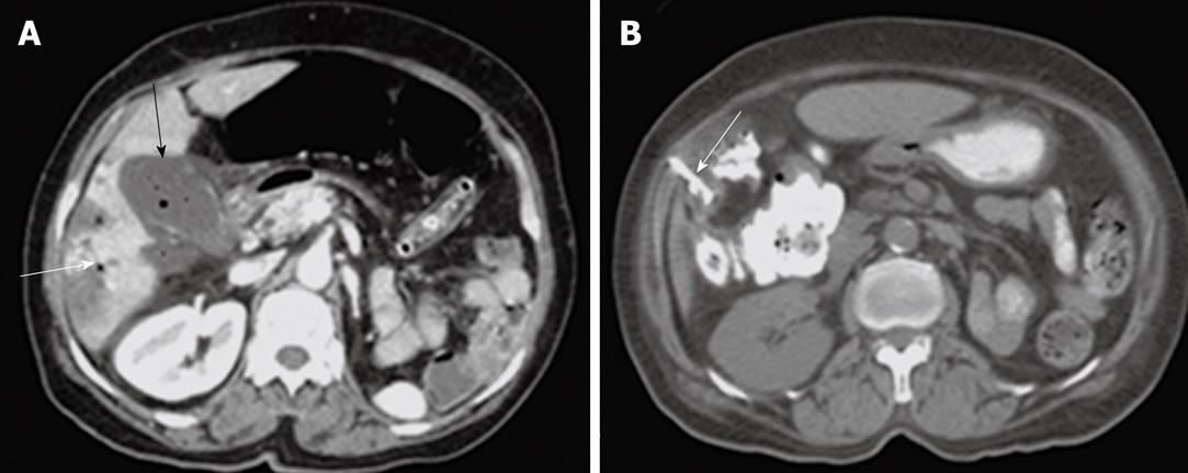
Figure 5 A case of emphysematous cholecystitis.
A: Post contrast computed tomography (CT) scan shows thick-enhancing walled gallbladder with turbid fluid content and air loculi seen within its lumen (black arrow). Minimal free pericholecystic fluid collection. Pneumobilia seen within the intrahepatic biliary radicles (white arrow); B: Follow up CT-guided cholecystostomy with drainage catheter seen within the gallbladder lumen (arrow).
- Citation: Donkol RH, Latif NA, Moghazy K. Percutaneous imaging-guided interventions for acute biliary disorders in high surgical risk patients. World J Radiol 2010; 2(9): 358-367
- URL: https://www.wjgnet.com/1949-8470/full/v2/i9/358.htm
- DOI: https://dx.doi.org/10.4329/wjr.v2.i9.358


