Copyright
©2010 Baishideng Publishing Group Co.
World J Radiol. Sep 28, 2010; 2(9): 358-367
Published online Sep 28, 2010. doi: 10.4329/wjr.v2.i9.358
Published online Sep 28, 2010. doi: 10.4329/wjr.v2.i9.358
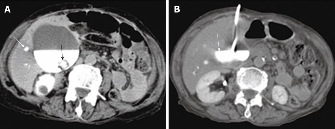
Figure 4 A case of gallbladder empyema.
A: Computed tomography (CT)-guided cholecystostomy shows opacification of a markedly dilated gallbladder with intraluminal filling defect due to the presence of gallstones (black arrow). The intrahepatic biliary ducts (white arrow) were opacified after endoscopic retrograde cholangiopancreatography, which failed to drain the biliary ducts; B: Follow up CT scan shows well-drained gallbladder (arrow).
- Citation: Donkol RH, Latif NA, Moghazy K. Percutaneous imaging-guided interventions for acute biliary disorders in high surgical risk patients. World J Radiol 2010; 2(9): 358-367
- URL: https://www.wjgnet.com/1949-8470/full/v2/i9/358.htm
- DOI: https://dx.doi.org/10.4329/wjr.v2.i9.358


