Copyright
©2010 Baishideng Publishing Group Co.
World J Radiol. Sep 28, 2010; 2(9): 345-353
Published online Sep 28, 2010. doi: 10.4329/wjr.v2.i9.345
Published online Sep 28, 2010. doi: 10.4329/wjr.v2.i9.345
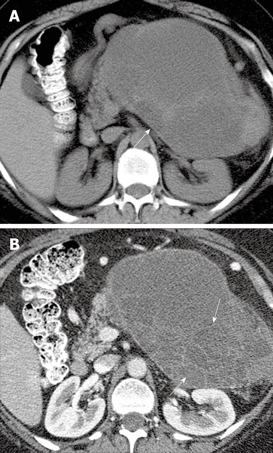
Figure 3 A 48-year-old woman with left upper quadrant pain.
A: Axial non-contrast computed tomography (CT) scan of the abdomen shows a heterogeneous mass (arrow) in the left upper quadrant; B: Axial contrast-enhanced CT of the abdomen shows enhancing septa (arrows) within the mass.
- Citation: Bhosale P, Balachandran A, Tamm E. Imaging of benign and malignant cystic pancreatic lesions and a strategy for follow up. World J Radiol 2010; 2(9): 345-353
- URL: https://www.wjgnet.com/1949-8470/full/v2/i9/345.htm
- DOI: https://dx.doi.org/10.4329/wjr.v2.i9.345


