Copyright
©2010 Baishideng Publishing Group Co.
World J Radiol. Sep 28, 2010; 2(9): 345-353
Published online Sep 28, 2010. doi: 10.4329/wjr.v2.i9.345
Published online Sep 28, 2010. doi: 10.4329/wjr.v2.i9.345
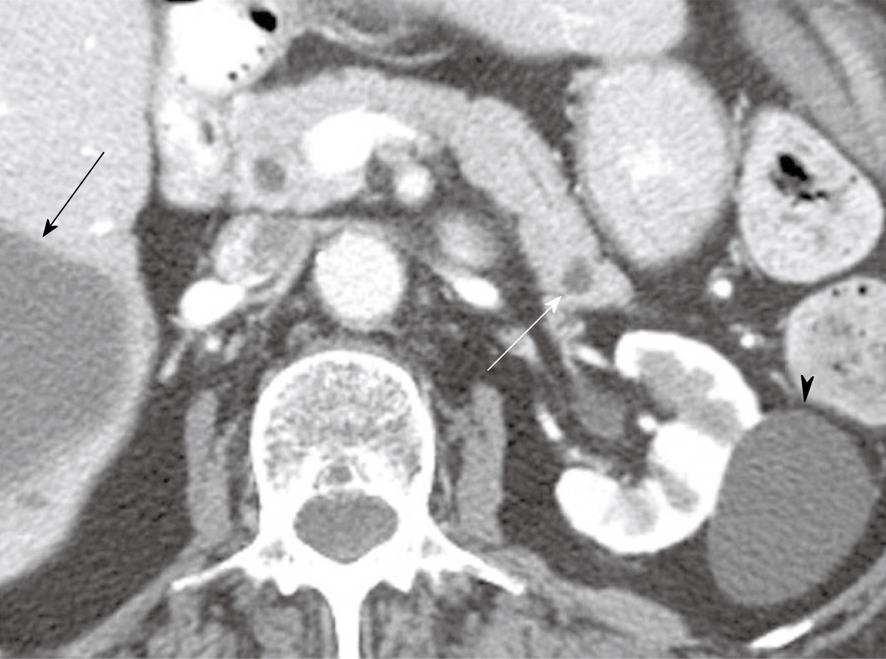
Figure 2 An 80-year-old woman with a history of hypertension, congestive heart failure and colon cancer.
Axial contrast-enhanced computed tomography of the abdomen shows a small low attenuation lesion in the pancreatic tail consistent with a cyst (white arrow), a cyst within the kidney (arrowhead) and a cyst within the liver (black arrow).
- Citation: Bhosale P, Balachandran A, Tamm E. Imaging of benign and malignant cystic pancreatic lesions and a strategy for follow up. World J Radiol 2010; 2(9): 345-353
- URL: https://www.wjgnet.com/1949-8470/full/v2/i9/345.htm
- DOI: https://dx.doi.org/10.4329/wjr.v2.i9.345


