Copyright
©2010 Baishideng Publishing Group Co.
World J Radiol. Aug 28, 2010; 2(8): 323-328
Published online Aug 28, 2010. doi: 10.4329/wjr.v2.i8.323
Published online Aug 28, 2010. doi: 10.4329/wjr.v2.i8.323
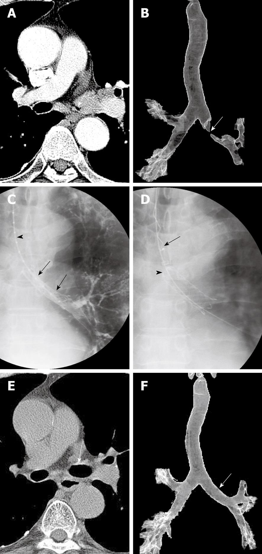
Figure 3 A 67-year-old man with a left main bronchial stricture caused by non-small-cell lung cancer.
A, B: Axial computed tomography (CT) scan (A) and anteroposterior view (B) of 3D surface-rendered reconstruction CT scan performed 3 d before stent placement, showing a severe left main bronchial stricture (arrows); C: Radiograph showing retrievable covered stent (arrows) 12 mm in diameter and 4 cm in length placed at the stricture. A sizing catheter (arrowhead) is also shown; D: Radiograph showing the collapse of the proximal end (arrowhead) of the stent while the retrievable hookwire (arrow) was withdrawn into the sheath; E, F: Axial CT scan (E) and anteroposterior view (F) of 3D surface-rendered reconstruction CT obtained 6 mo after stent removal, showing marked improvement in the stricture (arrows).
- Citation: Shin JH. Interventional management of tracheobronchial strictures. World J Radiol 2010; 2(8): 323-328
- URL: https://www.wjgnet.com/1949-8470/full/v2/i8/323.htm
- DOI: https://dx.doi.org/10.4329/wjr.v2.i8.323


