Copyright
©2010 Baishideng Publishing Group Co.
World J Radiol. Aug 28, 2010; 2(8): 323-328
Published online Aug 28, 2010. doi: 10.4329/wjr.v2.i8.323
Published online Aug 28, 2010. doi: 10.4329/wjr.v2.i8.323
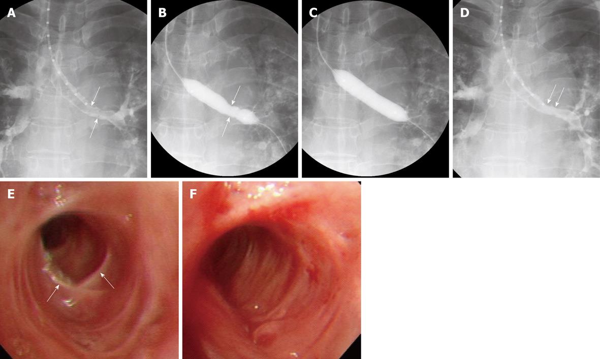
Figure 1 A 43-year-old woman with left main bronchial stricture caused by tuberculosis.
A: Radiograph shows irregular narrowing (arrows) of the left main bronchus; B, C: Radiographs show waist formation (arrows in B) of the inflated balloon and subsequent full inflation; D: Radiograph shows marked improvement in stricture (arrows); E, F: Bronchoscopic images before (E) and after (F) balloon dilation show substantial improvement in the stricture (arrows in E).
- Citation: Shin JH. Interventional management of tracheobronchial strictures. World J Radiol 2010; 2(8): 323-328
- URL: https://www.wjgnet.com/1949-8470/full/v2/i8/323.htm
- DOI: https://dx.doi.org/10.4329/wjr.v2.i8.323


