Copyright
©2010 Baishideng Publishing Group Co.
World J Radiol. Aug 28, 2010; 2(8): 309-322
Published online Aug 28, 2010. doi: 10.4329/wjr.v2.i8.309
Published online Aug 28, 2010. doi: 10.4329/wjr.v2.i8.309
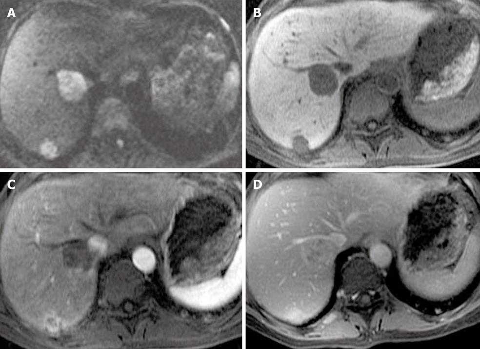
Figure 9 A 64-year-old with liver metastases from a primary sarcoma.
A: The diffusion weighted imaging at b = 500 shows two solid lesions in the right lobe of the liver; B-D: The pre-contrast (B), late arterial (C), and delayed phase (D) show late enhancement. The enhancement pattern is similar to intrahepatic cholangiocarcinoma.
- Citation: Maniam S, Szklaruk J. Magnetic resonance imaging: Review of imaging techniques and overview of liver imaging. World J Radiol 2010; 2(8): 309-322
- URL: https://www.wjgnet.com/1949-8470/full/v2/i8/309.htm
- DOI: https://dx.doi.org/10.4329/wjr.v2.i8.309


