Copyright
©2010 Baishideng Publishing Group Co.
World J Radiol. Aug 28, 2010; 2(8): 298-308
Published online Aug 28, 2010. doi: 10.4329/wjr.v2.i8.298
Published online Aug 28, 2010. doi: 10.4329/wjr.v2.i8.298
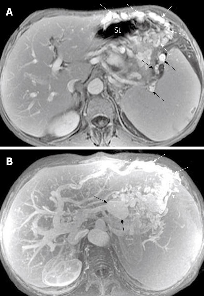
Figure 20 Pancreatic regional portal hypertension in a 36-year-old man with a history of acute pancreatitis.
A: Axial T1-weighted image obtained with intravenous contrast material reveals enhancement of numerous and circuitous veins (white arrows) around the gastric fundus, and splenic veins (black arrows) adjacent to the splenic hilum; B: Magnetic resonance angiography depicts the establishment of numerous and conspicuous collateral vessels due to gastric fundic varices (white arrows) and a pseudoaneurysm (black arrows). St: Stomach.
- Citation: Xiao B, Zhang XM. Magnetic resonance imaging for acute pancreatitis. World J Radiol 2010; 2(8): 298-308
- URL: https://www.wjgnet.com/1949-8470/full/v2/i8/298.htm
- DOI: https://dx.doi.org/10.4329/wjr.v2.i8.298


