Copyright
©2010 Baishideng Publishing Group Co.
World J Radiol. Aug 28, 2010; 2(8): 298-308
Published online Aug 28, 2010. doi: 10.4329/wjr.v2.i8.298
Published online Aug 28, 2010. doi: 10.4329/wjr.v2.i8.298
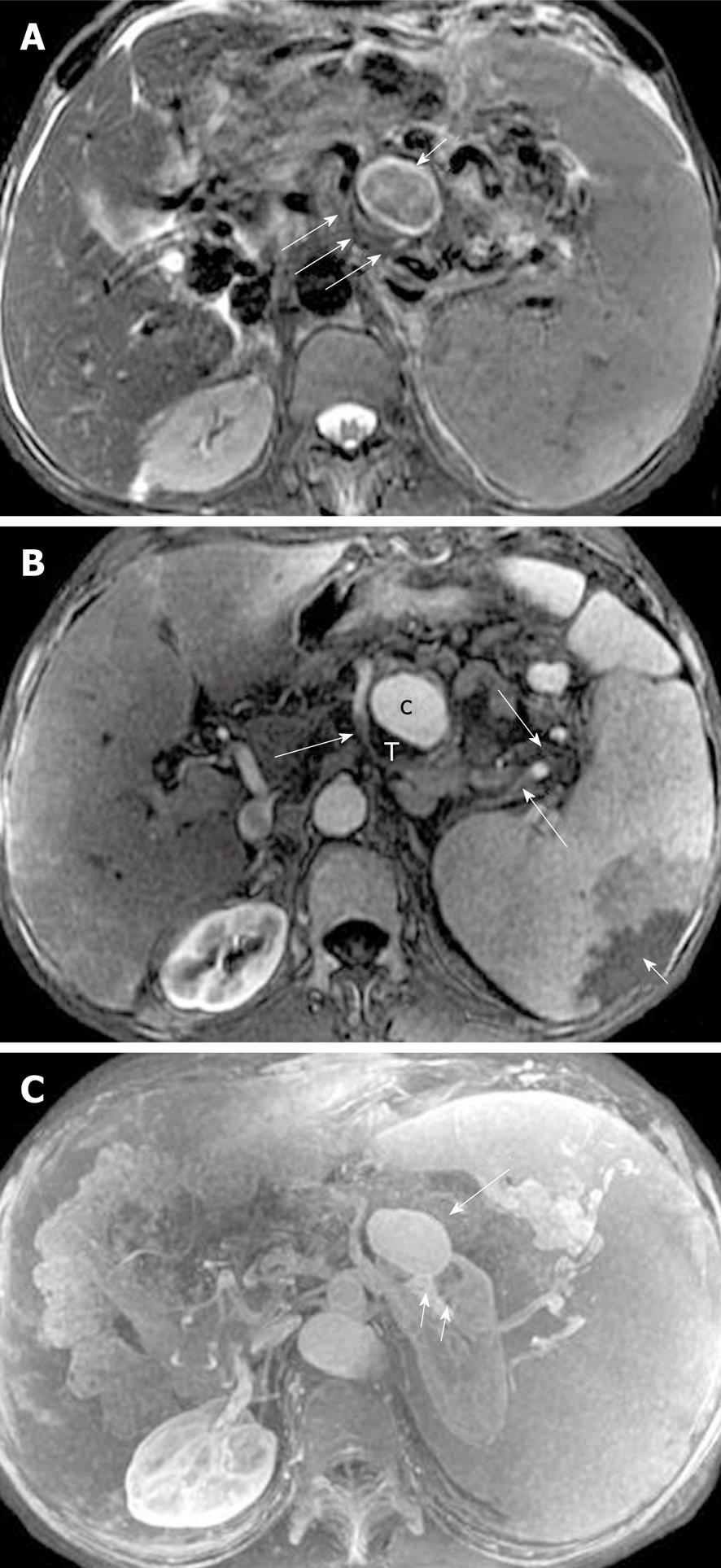
Figure 18 Splenic artery pseudoaneurysm in a 36-year-old man with a history of acute pancreatitis.
A: Axial magnetic resonance (MR) T2-weighted with fat-suppression image shows the involved part of the splenic artery (large arrows) and aneurysmal dilatation (small arrows); B: Axial T1-weighted image obtained with intravenous contrast material reveals enhancement of the pseudoaneurysm cavity (c) and a filling defect present as mural thrombosis (T), and wedge-shape zones of infarction of the spleen (small arrows) due to the involved parts of the splenic artery (large arrows); C: MR angiography further depicts the visualization of the relationship between this pseudoaneurysm (large arrow) and the involved splenic artery (small arrows). c: Cavity of the pseudoaneurysm; T: Mural thrombosis in the pseudoaneurysm.
- Citation: Xiao B, Zhang XM. Magnetic resonance imaging for acute pancreatitis. World J Radiol 2010; 2(8): 298-308
- URL: https://www.wjgnet.com/1949-8470/full/v2/i8/298.htm
- DOI: https://dx.doi.org/10.4329/wjr.v2.i8.298


