Copyright
©2010 Baishideng Publishing Group Co.
World J Radiol. Aug 28, 2010; 2(8): 298-308
Published online Aug 28, 2010. doi: 10.4329/wjr.v2.i8.298
Published online Aug 28, 2010. doi: 10.4329/wjr.v2.i8.298
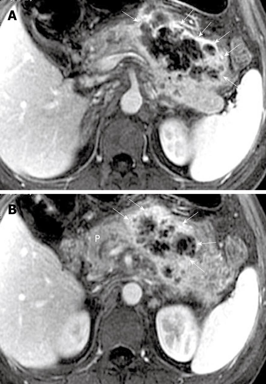
Figure 15 Acute necrotic pancreatitis and peripancreatic cellulitis in a 39-year-old man.
A, B: Axial magnetic resonance T1-weighted images obtained with intravenous contrast material reveal an ill-defined, multilocular, inflammatory mass (arrows) with ring-like and separated enhancement adjacent to the body and tail of the pancreas. P: Pancreas.
- Citation: Xiao B, Zhang XM. Magnetic resonance imaging for acute pancreatitis. World J Radiol 2010; 2(8): 298-308
- URL: https://www.wjgnet.com/1949-8470/full/v2/i8/298.htm
- DOI: https://dx.doi.org/10.4329/wjr.v2.i8.298


