Copyright
©2010 Baishideng Publishing Group Co.
World J Radiol. Aug 28, 2010; 2(8): 298-308
Published online Aug 28, 2010. doi: 10.4329/wjr.v2.i8.298
Published online Aug 28, 2010. doi: 10.4329/wjr.v2.i8.298
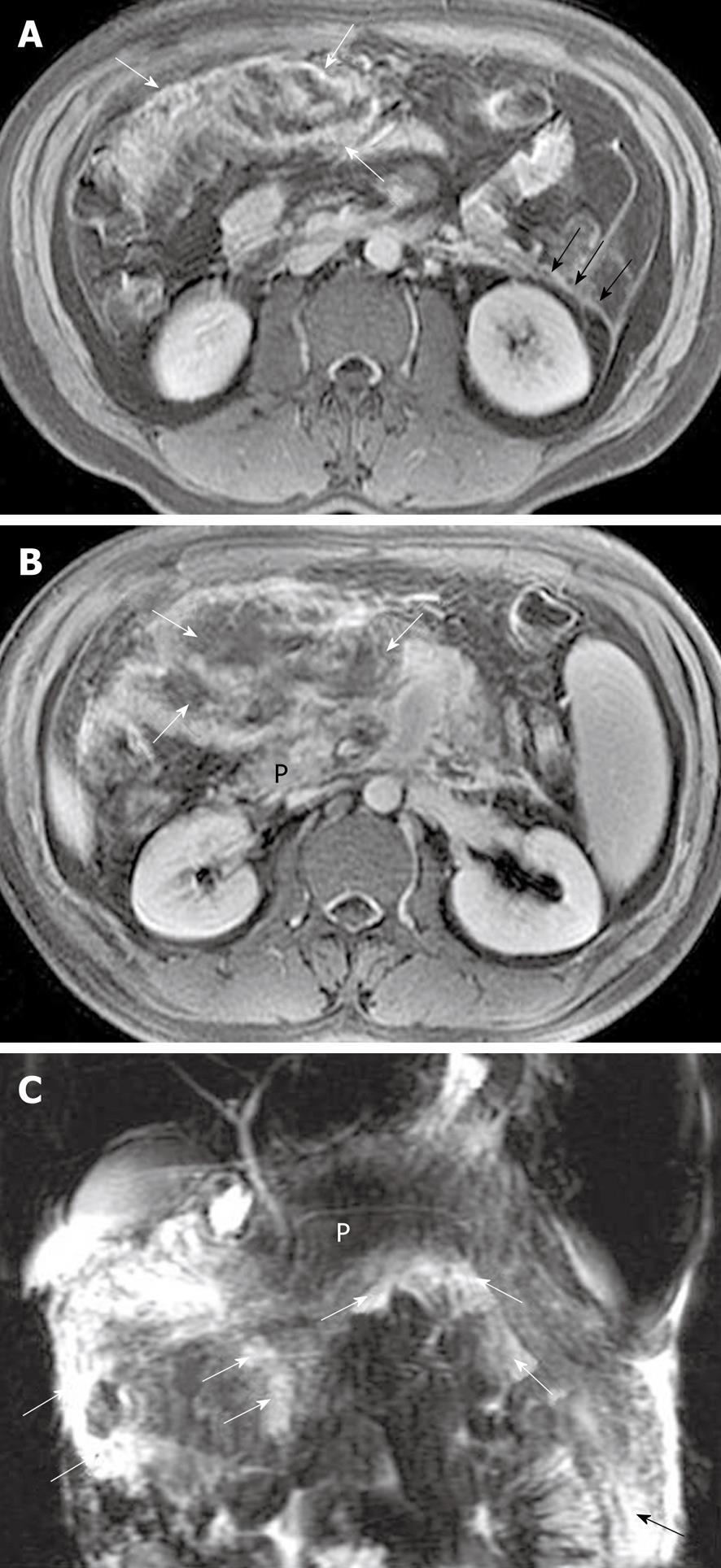
Figure 12 Pancreatic focal necrosis in a 35-year-old man after an episode of acute pancreatitis.
A, B: Enhanced axial T1-weighted images reveal the irregular thickening and heterogeneous enhancement of the intestinal wall (white arrows in A), anterior fascia of the left kidney (black arrows in A), and mesenteric edema associated with fat necrosis (arrows in B); C: Magnetic resonance cholangiopancreatography reveals multiple edema and small fluid collections (arrows) adjacent to small intestine and colon. P: Pancreas.
- Citation: Xiao B, Zhang XM. Magnetic resonance imaging for acute pancreatitis. World J Radiol 2010; 2(8): 298-308
- URL: https://www.wjgnet.com/1949-8470/full/v2/i8/298.htm
- DOI: https://dx.doi.org/10.4329/wjr.v2.i8.298


