Copyright
©2010 Baishideng Publishing Group Co.
World J Radiol. Aug 28, 2010; 2(8): 298-308
Published online Aug 28, 2010. doi: 10.4329/wjr.v2.i8.298
Published online Aug 28, 2010. doi: 10.4329/wjr.v2.i8.298
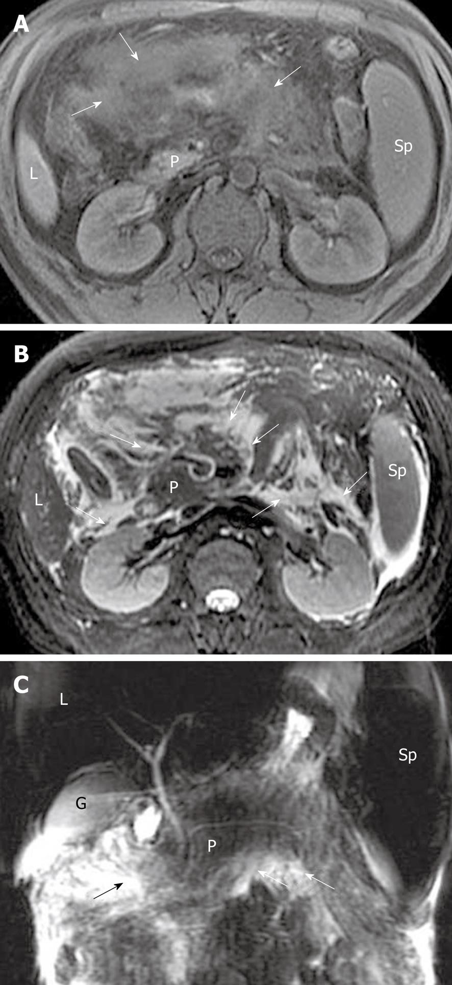
Figure 10 Acute pancreatitis in a 34-year-old man.
Axial magnetic resonance (MR) T1-weighted with fat-suppression (A) and T2-weighted with fat-suppression (B) images reveal peripancreatic inflammation extension co-existing with fat necrosis (arrows) in mesenteric fat tissue regions and anterior pararenal space of both kidneys. Extravasated fluid (arrows) is also present around the gland on MR cholangiopancreatography (C). L: Liver; P: Pancreas; Sp: Spleen; G: Gallbladder.
- Citation: Xiao B, Zhang XM. Magnetic resonance imaging for acute pancreatitis. World J Radiol 2010; 2(8): 298-308
- URL: https://www.wjgnet.com/1949-8470/full/v2/i8/298.htm
- DOI: https://dx.doi.org/10.4329/wjr.v2.i8.298


