Copyright
©2010 Baishideng Publishing Group Co.
World J Radiol. Aug 28, 2010; 2(8): 298-308
Published online Aug 28, 2010. doi: 10.4329/wjr.v2.i8.298
Published online Aug 28, 2010. doi: 10.4329/wjr.v2.i8.298
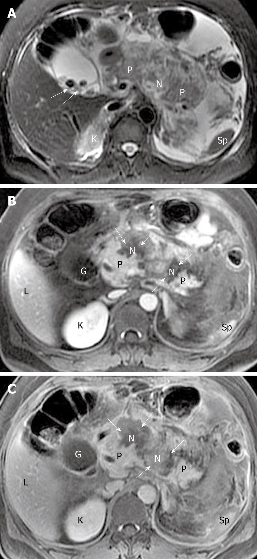
Figure 6 Gallstones, acute pancreatitis, and gland liquefied necrosis in a 33-year-old woman.
Axial magnetic resonance T2-weighted with fat-suppression image (A) shows hypointense gallstones (arrows), and axial T1-weighted images obtained in late arterial phase (B) and venous phase (C) reveal two zones (arrows) of pancreatic liquefied necrosis in the neck and body of the gland (like “rupture of the pancreas”). The extent of necrosis is > 50% of the pancreatic gland. The head and the tail of the pancreas are still enhancing (P). N: Liquefied gland necrosis; G: Gallbladder; K: Kidney; L: Liver; P: Pancreas; Sp: Spleen.
- Citation: Xiao B, Zhang XM. Magnetic resonance imaging for acute pancreatitis. World J Radiol 2010; 2(8): 298-308
- URL: https://www.wjgnet.com/1949-8470/full/v2/i8/298.htm
- DOI: https://dx.doi.org/10.4329/wjr.v2.i8.298


