Copyright
©2010 Baishideng Publishing Group Co.
World J Radiol. Aug 28, 2010; 2(8): 298-308
Published online Aug 28, 2010. doi: 10.4329/wjr.v2.i8.298
Published online Aug 28, 2010. doi: 10.4329/wjr.v2.i8.298
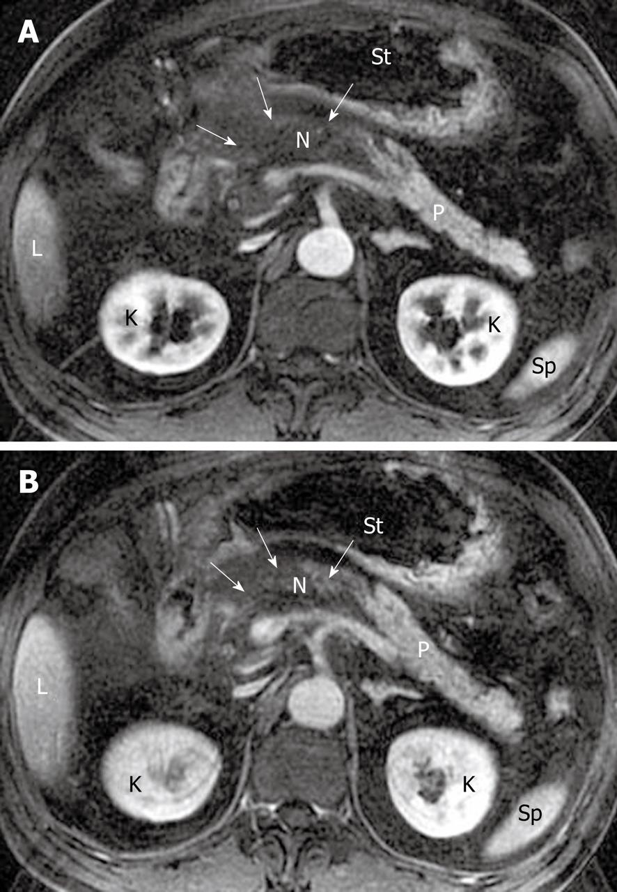
Figure 5 Pancreatic diffuse necrosis in a 65-year-old man after an episode of acute pancreatitis.
A, B: Axial magnetic resonance T1-weighted images obtained in late arterial phase and venous phase reveal large necrotic areas (arrows) (“black pancreas”) in the pancreatic head, neck and part of the body. The extent of necrosis is up to 30-50% of the pancreatic gland. N: Necrosis; K: Kidney; L: Liver; P: Pancreas; Sp: Spleen; St: Stomach.
- Citation: Xiao B, Zhang XM. Magnetic resonance imaging for acute pancreatitis. World J Radiol 2010; 2(8): 298-308
- URL: https://www.wjgnet.com/1949-8470/full/v2/i8/298.htm
- DOI: https://dx.doi.org/10.4329/wjr.v2.i8.298


