Copyright
©2010 Baishideng Publishing Group Co.
World J Radiol. Aug 28, 2010; 2(8): 298-308
Published online Aug 28, 2010. doi: 10.4329/wjr.v2.i8.298
Published online Aug 28, 2010. doi: 10.4329/wjr.v2.i8.298
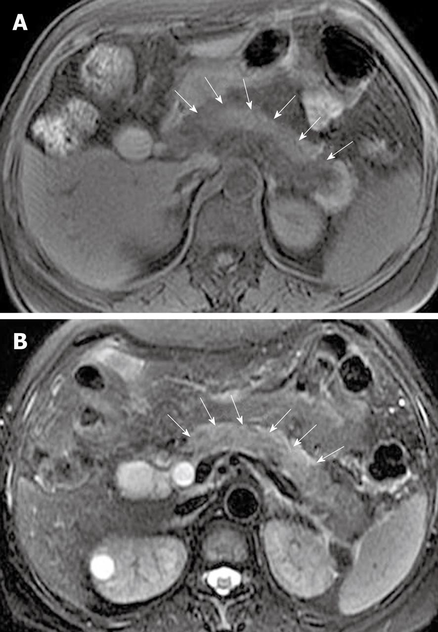
Figure 2 Acute edematous pancreatitis in a 29-year-old man.
Axial non-enhanced magnetic resonance T1-weighted with fat-suppression image (A) and axial T2-weighted with fat-suppression image (B) show that the parenchyma of the pancreatic head, body and part of the tail is hypointense (arrows in A) and hyperintense (arrows in B) relative to the liver.
- Citation: Xiao B, Zhang XM. Magnetic resonance imaging for acute pancreatitis. World J Radiol 2010; 2(8): 298-308
- URL: https://www.wjgnet.com/1949-8470/full/v2/i8/298.htm
- DOI: https://dx.doi.org/10.4329/wjr.v2.i8.298


