Copyright
©2010 Baishideng Publishing Group Co.
World J Radiol. Jul 28, 2010; 2(7): 283-288
Published online Jul 28, 2010. doi: 10.4329/wjr.v2.i7.283
Published online Jul 28, 2010. doi: 10.4329/wjr.v2.i7.283
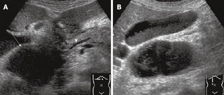
Figure 2 Abdominal ultrasonography showed a 65 mm × 45 mm heterogeneous mass at the level of the pancreatic head (arrows) (A and B), with a slightly dilated main pancreatic duct (arrowhead) and peripancreatic fluid collection (A).
- Citation: Shiozawa K, Watanabe M, Igarashi Y, Matsukiyo Y, Matsui T, Sumino Y. Acute pancreatitis secondary to intramural duodenal hematoma: Case report and literature review. World J Radiol 2010; 2(7): 283-288
- URL: https://www.wjgnet.com/1949-8470/full/v2/i7/283.htm
- DOI: https://dx.doi.org/10.4329/wjr.v2.i7.283


