Copyright
©2010 Baishideng Publishing Group Co.
World J Radiol. Jul 28, 2010; 2(7): 283-288
Published online Jul 28, 2010. doi: 10.4329/wjr.v2.i7.283
Published online Jul 28, 2010. doi: 10.4329/wjr.v2.i7.283
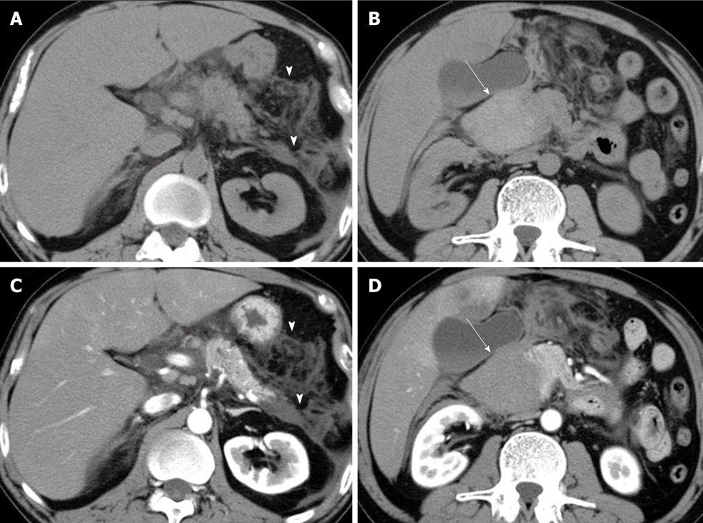
Figure 1 Abdominal computed tomography revealed an increased density of peripancreatic dirty fat tissue and fluid collection (arrowheads), without (A) and with (C) contrast.
Unenhanced computed tomography (B) revealed a slightly hyperdense, spindle-shaped mass, 65 mm in maximum diameter, mainly along the descending part of the duodenum (arrow). This mass showed no contrast enhancement in the arterial phase (arrow) (D).
- Citation: Shiozawa K, Watanabe M, Igarashi Y, Matsukiyo Y, Matsui T, Sumino Y. Acute pancreatitis secondary to intramural duodenal hematoma: Case report and literature review. World J Radiol 2010; 2(7): 283-288
- URL: https://www.wjgnet.com/1949-8470/full/v2/i7/283.htm
- DOI: https://dx.doi.org/10.4329/wjr.v2.i7.283


