Copyright
©2010 Baishideng Publishing Group Co.
World J Radiol. Jul 28, 2010; 2(7): 249-256
Published online Jul 28, 2010. doi: 10.4329/wjr.v2.i7.249
Published online Jul 28, 2010. doi: 10.4329/wjr.v2.i7.249
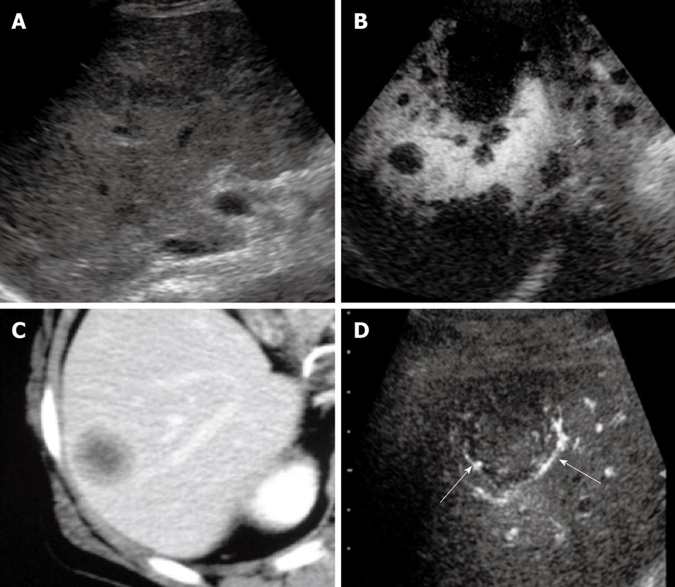
Figure 5 Liver metastasis.
A: Multiple masses were seen in the liver by B-mode ultrasonography (US); B: Multiple defects were seen by Sonazoid-enhanced US in the post-vascular phase; C: Portal phase dynamic scan detected a hypoenhanced nodule in segment 6 of the liver; D: Peripheral enhancement of the nodule (arrows) was obtained by Sonazoid-enhanced ultrasonography in the early vascular phase.
- Citation: Minami Y, Kudo M. Hepatic malignancies: Correlation between sonographic findings and pathological features. World J Radiol 2010; 2(7): 249-256
- URL: https://www.wjgnet.com/1949-8470/full/v2/i7/249.htm
- DOI: https://dx.doi.org/10.4329/wjr.v2.i7.249


