Copyright
©2010 Baishideng Publishing Group Co.
World J Radiol. Jul 28, 2010; 2(7): 249-256
Published online Jul 28, 2010. doi: 10.4329/wjr.v2.i7.249
Published online Jul 28, 2010. doi: 10.4329/wjr.v2.i7.249
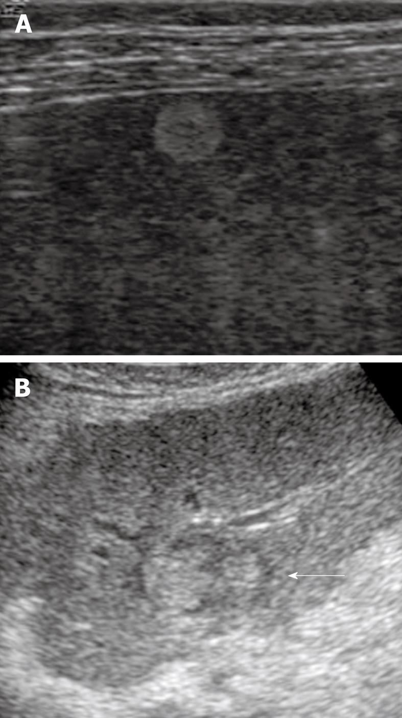
Figure 4 Early hepatocellular carcinoma.
A: A nodule that was 1.5 cm in diameter in segment 5 of the liver was shown as highly echoic because of fatty changes in the nodule; B: A nodule-in-nodule appearance (arrow) was demonstrated as a hyperechoic tumor within a hypoechoic nodule.
- Citation: Minami Y, Kudo M. Hepatic malignancies: Correlation between sonographic findings and pathological features. World J Radiol 2010; 2(7): 249-256
- URL: https://www.wjgnet.com/1949-8470/full/v2/i7/249.htm
- DOI: https://dx.doi.org/10.4329/wjr.v2.i7.249


