Copyright
©2010 Baishideng Publishing Group Co.
World J Radiol. Jul 28, 2010; 2(7): 237-248
Published online Jul 28, 2010. doi: 10.4329/wjr.v2.i7.237
Published online Jul 28, 2010. doi: 10.4329/wjr.v2.i7.237
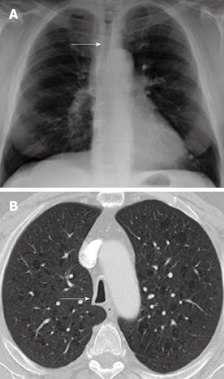
Figure 6 Saber sheath trachea.
A: Chest radiograph PA view demonstrating diffuse narrowing of the trachea (arrow); B: Axial computed tomography scan showing inward bowing of the lateral tracheal wall (arrow) with elongated sagittal dimension of the trachea compared to the coronal plane is consistent with the saber sheath configuration.
- Citation: Laroia AT, Thompson BH, Laroia ST, Beek EJV. Modern imaging of the tracheo-bronchial tree. World J Radiol 2010; 2(7): 237-248
- URL: https://www.wjgnet.com/1949-8470/full/v2/i7/237.htm
- DOI: https://dx.doi.org/10.4329/wjr.v2.i7.237


