Copyright
©2010 Baishideng Publishing Group Co.
World J Radiol. Jul 28, 2010; 2(7): 237-248
Published online Jul 28, 2010. doi: 10.4329/wjr.v2.i7.237
Published online Jul 28, 2010. doi: 10.4329/wjr.v2.i7.237
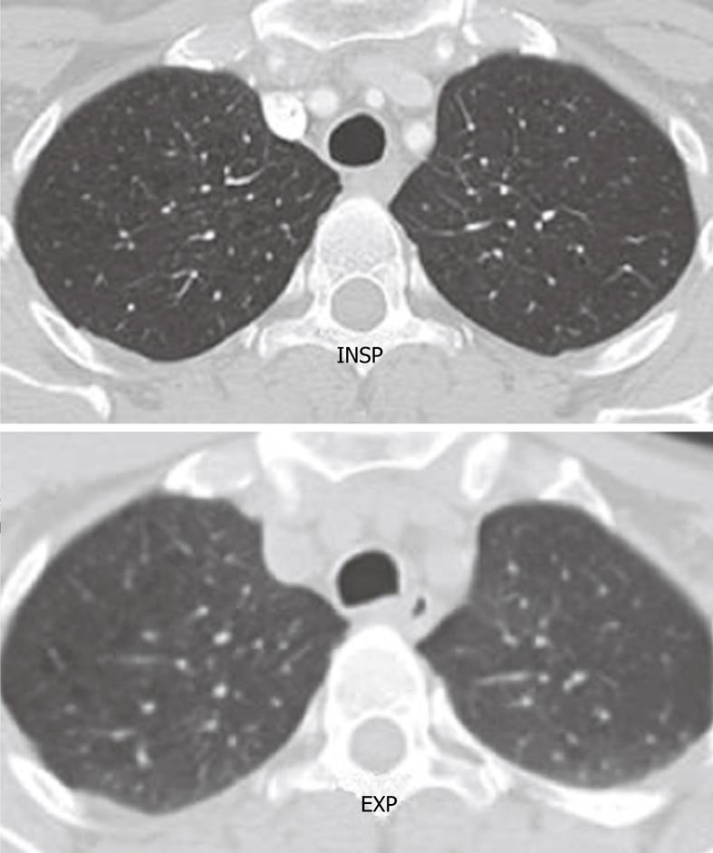
Figure 2 Axial computed tomography image shows the normal rounded configuration of the trachea at the end of inspiration.
Note the normal anterior bowing of the posterior membranous wall of the trachea at the end of expiration.
- Citation: Laroia AT, Thompson BH, Laroia ST, Beek EJV. Modern imaging of the tracheo-bronchial tree. World J Radiol 2010; 2(7): 237-248
- URL: https://www.wjgnet.com/1949-8470/full/v2/i7/237.htm
- DOI: https://dx.doi.org/10.4329/wjr.v2.i7.237


