Copyright
©2010 Baishideng Publishing Group Co.
World J Radiol. Jun 28, 2010; 2(6): 224-229
Published online Jun 28, 2010. doi: 10.4329/wjr.v2.i6.224
Published online Jun 28, 2010. doi: 10.4329/wjr.v2.i6.224
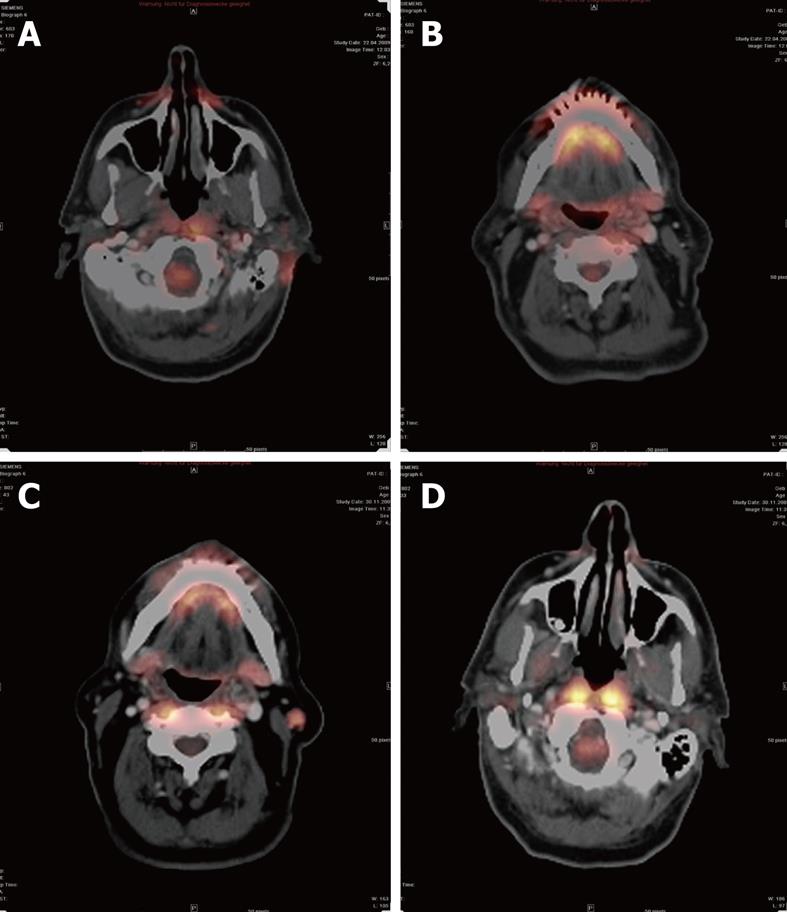
Figure 3 Positron emission tomography-CT after resection of a melanocytic tumor of uncertain malignant origin.
A: The resection area still showed a higher glucose metabolism than normal healthy tissue at 2 mo after therapy; B: A small lymph node (LN), but no sign of higher metabolism; C: A malignant LN with higher metabolism; D: There was no higher glucose metabolism noticeable at 6 mo after therapy.
- Citation: Vogl TJ, Harth M, Siebenhandl P. Different imaging techniques in the head and neck: Assets and drawbacks. World J Radiol 2010; 2(6): 224-229
- URL: https://www.wjgnet.com/1949-8470/full/v2/i6/224.htm
- DOI: https://dx.doi.org/10.4329/wjr.v2.i6.224


