Copyright
©2010 Baishideng Publishing Group Co.
World J Radiol. Jun 28, 2010; 2(6): 215-223
Published online Jun 28, 2010. doi: 10.4329/wjr.v2.i6.215
Published online Jun 28, 2010. doi: 10.4329/wjr.v2.i6.215
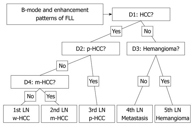
Figure 2 Illustration of the decision tree model used in this study.
Four decision nodes in which alternative choice was determined by all five FLLs, leading to a final diagnostic decision for five liver lesions. D: Decision node; FLL: Focal liver lesion; HCC: Hepatocellular carcinoma; LN: Leaf node; m-HCC: Moderately differentiated HCC; p-HCC: Poorly differentiated HCC; w-HCC: Well-differentiated HCC.
- Citation: Sugimoto K, Shiraishi J, Moriyasu F, Doi K. Computer-aided diagnosis for contrast-enhanced ultrasound in the liver. World J Radiol 2010; 2(6): 215-223
- URL: https://www.wjgnet.com/1949-8470/full/v2/i6/215.htm
- DOI: https://dx.doi.org/10.4329/wjr.v2.i6.215


