Copyright
©2010 Baishideng Publishing Group Co.
World J Radiol. Jun 28, 2010; 2(6): 203-214
Published online Jun 28, 2010. doi: 10.4329/wjr.v2.i6.203
Published online Jun 28, 2010. doi: 10.4329/wjr.v2.i6.203
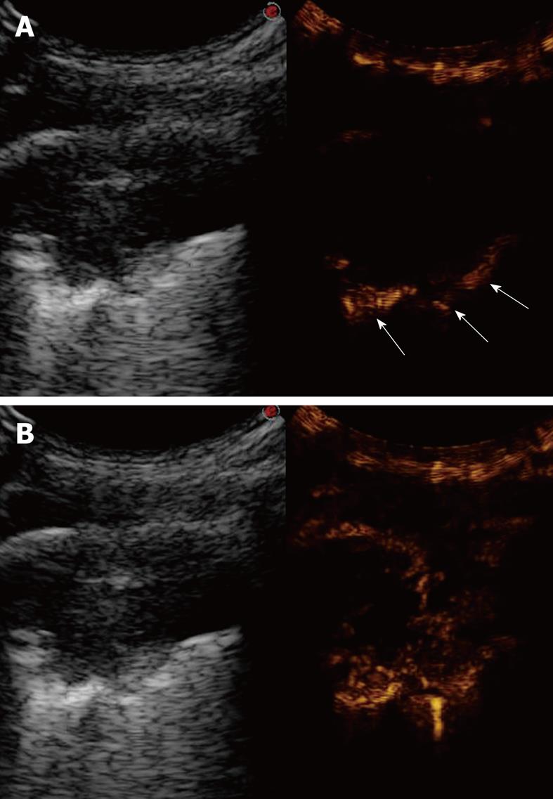
Figure 19 Contrast-enhanced ultrasonography of bronchial carcinoma.
A: Baseline scan shows a hypoechoic lesion with irregular borders (left side of the split-screen). Ten seconds after iv bolus of contrast agent, the pulmonary parenchyma near the lesion is already enhanced (arrows), whereas the lesions is still unenhanced (right side of the split-screen); B: Twenty seconds later, the lesion shows delayed inhomogeneous enhancement, which indicates a preferential bronchial arterial supply (right side of the split-screen).
- Citation: Sartori S, Tombesi P. Emerging roles for transthoracic ultrasonography in pulmonary diseases. World J Radiol 2010; 2(6): 203-214
- URL: https://www.wjgnet.com/1949-8470/full/v2/i6/203.htm
- DOI: https://dx.doi.org/10.4329/wjr.v2.i6.203


