Copyright
©2010 Baishideng Publishing Group Co.
World J Radiol. Jun 28, 2010; 2(6): 203-214
Published online Jun 28, 2010. doi: 10.4329/wjr.v2.i6.203
Published online Jun 28, 2010. doi: 10.4329/wjr.v2.i6.203
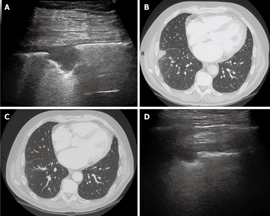
Figure 11 Dynamic course of pulmonary infarction.
A: Lateral intercostal scan of the right lung shows a typical triangular-shaped peripheral lesion; B: Likewise, computed tomography scan of the lateral segment of the lower right lobe shows a triangular pleural-based lesion with the vertex towards the hilum; C: After 40 d, the lesion is no longer visible by computed tomography scan; D: The lesion appears reduced in size at transthoracic ultrasonography examination.
- Citation: Sartori S, Tombesi P. Emerging roles for transthoracic ultrasonography in pulmonary diseases. World J Radiol 2010; 2(6): 203-214
- URL: https://www.wjgnet.com/1949-8470/full/v2/i6/203.htm
- DOI: https://dx.doi.org/10.4329/wjr.v2.i6.203


