Copyright
©2010 Baishideng Publishing Group Co.
World J Radiol. Jun 28, 2010; 2(6): 203-214
Published online Jun 28, 2010. doi: 10.4329/wjr.v2.i6.203
Published online Jun 28, 2010. doi: 10.4329/wjr.v2.i6.203
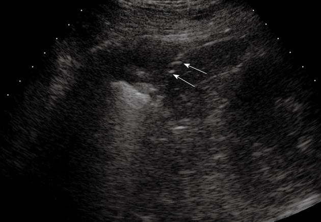
Figure 10 Pulmonary infarction.
Posterior intercostal scan shows a triangular-shaped hypoechoic lesion with central hyperechoic structures that indicate the presence of air occupying the affected bronchiole (arrows).
- Citation: Sartori S, Tombesi P. Emerging roles for transthoracic ultrasonography in pulmonary diseases. World J Radiol 2010; 2(6): 203-214
- URL: https://www.wjgnet.com/1949-8470/full/v2/i6/203.htm
- DOI: https://dx.doi.org/10.4329/wjr.v2.i6.203


