Copyright
©2010 Baishideng Publishing Group Co.
World J Radiol. May 28, 2010; 2(5): 159-165
Published online May 28, 2010. doi: 10.4329/wjr.v2.i5.159
Published online May 28, 2010. doi: 10.4329/wjr.v2.i5.159
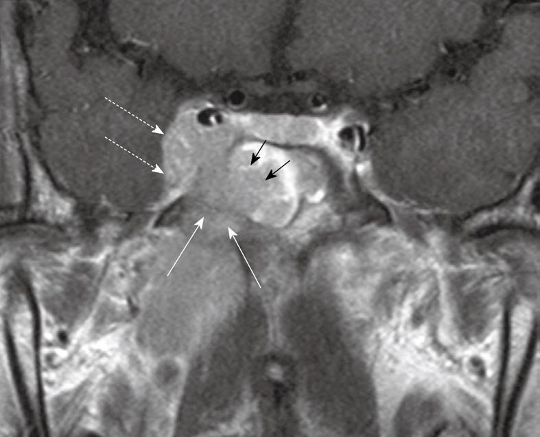
Figure 9 Coronal post-contrast T1 weighted MRI showing a nasopharyngeal carcinoma with direct infiltration through the sphenoid body (long arrows) into the sphenoid sinus (short arrows) and right cavernous sinus (broken arrows).
- Citation: King AD, Bhatia KSS. Magnetic resonance imaging staging of nasopharyngeal carcinoma in the head and neck. World J Radiol 2010; 2(5): 159-165
- URL: https://www.wjgnet.com/1949-8470/full/v2/i5/159.htm
- DOI: https://dx.doi.org/10.4329/wjr.v2.i5.159


