Copyright
©2010 Baishideng Publishing Group Co.
World J Radiol. May 28, 2010; 2(5): 151-158
Published online May 28, 2010. doi: 10.4329/wjr.v2.i5.151
Published online May 28, 2010. doi: 10.4329/wjr.v2.i5.151
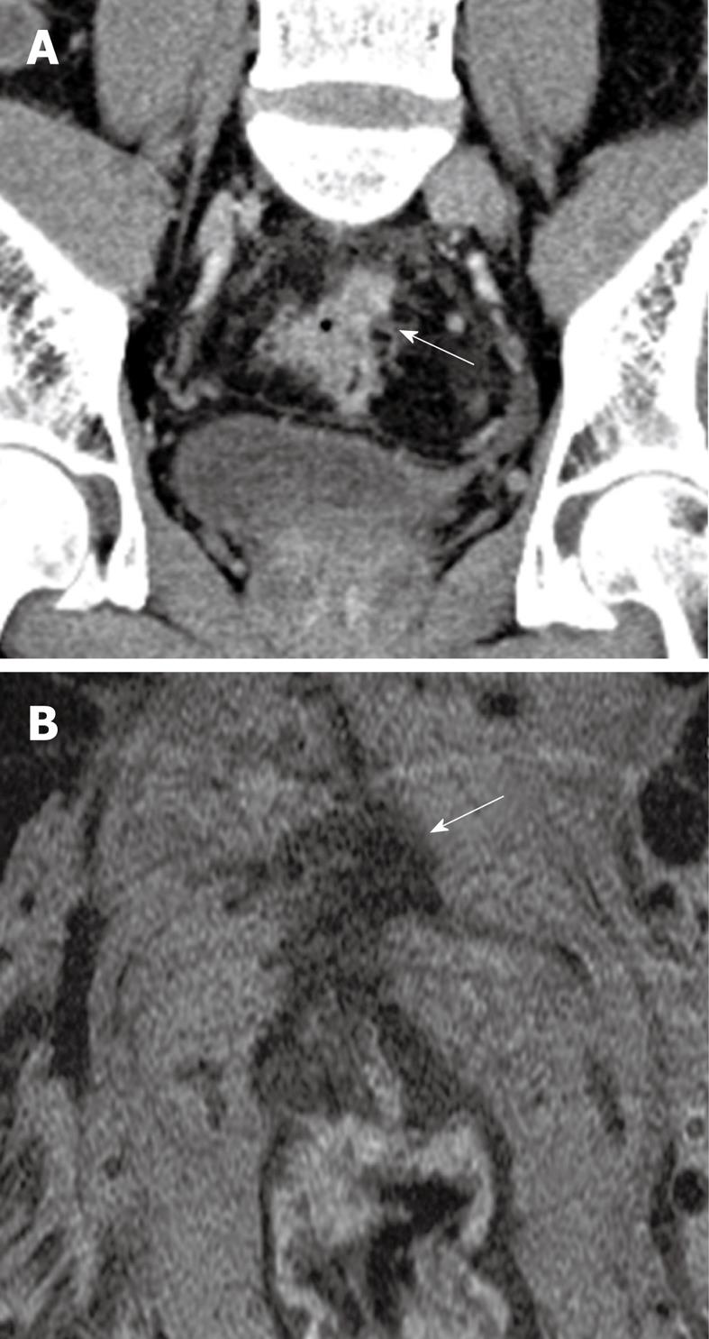
Figure 2 53-year-old male patient.
Presented with lower gastrointestinal bleeding. Endoscopy revealed a fungating mass in the proximal rectum, preventing passage of the scope more proximally. A: Coronal reconstructed contrast-enhanced CT image reveals a spiculated mass at the rectosigmoid junction with a spiculated extraserosal nodular component (arrow); B: Corresponding T2 weighted high resolution magnetic resonance image in the axial plane confirms the findings of extraserosal extension of disease (arrow). The patient was referred for assessment of suitability for neoadjuvant chemoradiation treatment.
- Citation: Tan CH, Iyer R. Use of computed tomography in the management of colorectal cancer. World J Radiol 2010; 2(5): 151-158
- URL: https://www.wjgnet.com/1949-8470/full/v2/i5/151.htm
- DOI: https://dx.doi.org/10.4329/wjr.v2.i5.151


