Copyright
©2010 Baishideng Publishing Group Co.
World J Radiol. May 28, 2010; 2(5): 151-158
Published online May 28, 2010. doi: 10.4329/wjr.v2.i5.151
Published online May 28, 2010. doi: 10.4329/wjr.v2.i5.151
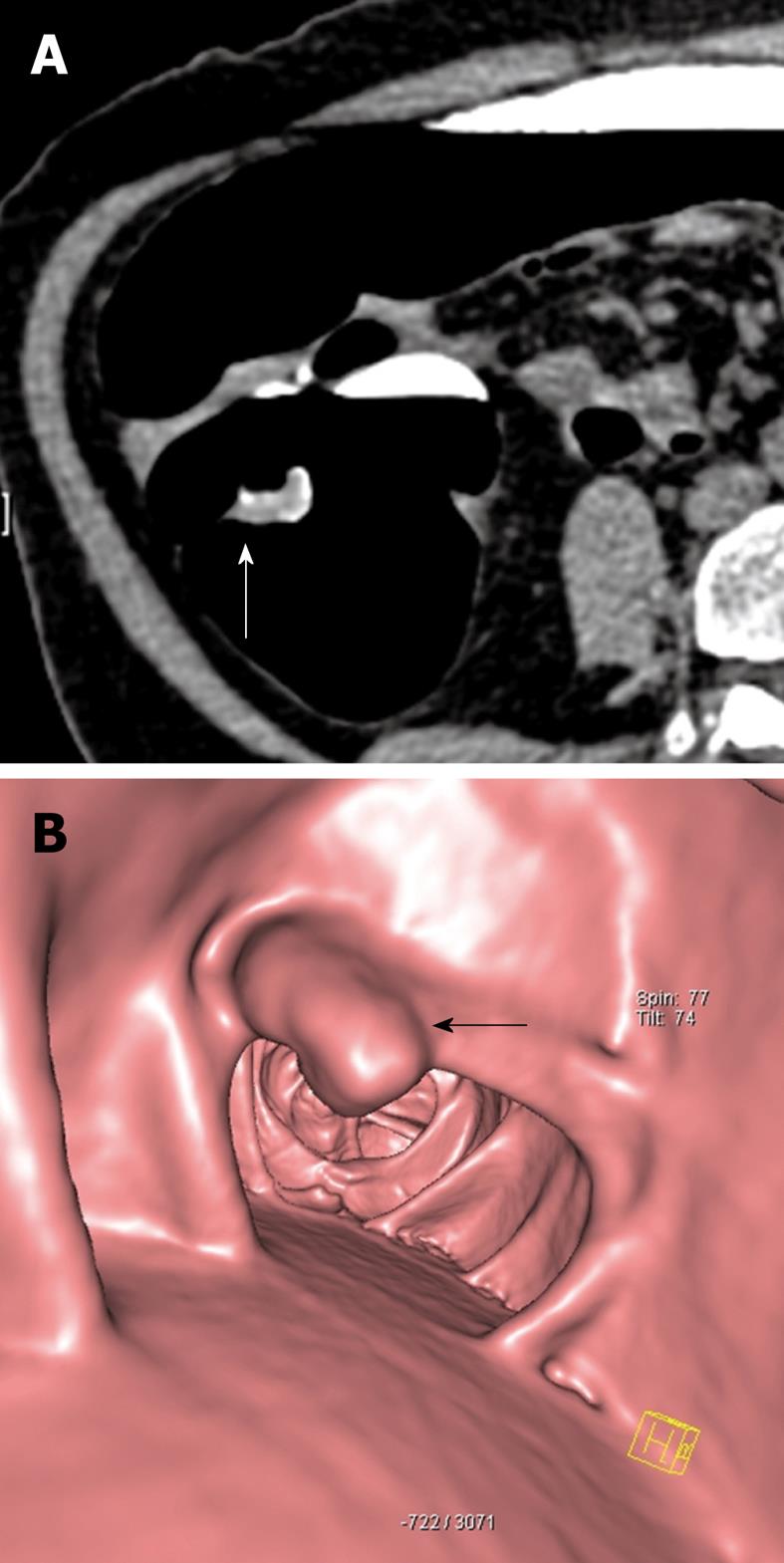
Figure 1 73-year-old female.
Faecal occult blood positive. Sigmoidoscopy was normal. A: Source axial computed tomography (CT) image from CT colonography study in the prone position demonstrates focal thickening (arrow) along a haustral fold in the proximal colon. Note the presence of contrast tagged faecal material coating the lesion; B: 3D reconstructed image of the same lesion showing a polypoid mass (arrow) arising from the haustral fold. Biopsy was positive for adenocarcinoma and the patient underwent curative right hemicolectomy.
- Citation: Tan CH, Iyer R. Use of computed tomography in the management of colorectal cancer. World J Radiol 2010; 2(5): 151-158
- URL: https://www.wjgnet.com/1949-8470/full/v2/i5/151.htm
- DOI: https://dx.doi.org/10.4329/wjr.v2.i5.151


