Copyright
©2010 Baishideng Publishing Group Co.
World J Radiol. Apr 28, 2010; 2(4): 143-147
Published online Apr 28, 2010. doi: 10.4329/wjr.v2.i4.143
Published online Apr 28, 2010. doi: 10.4329/wjr.v2.i4.143
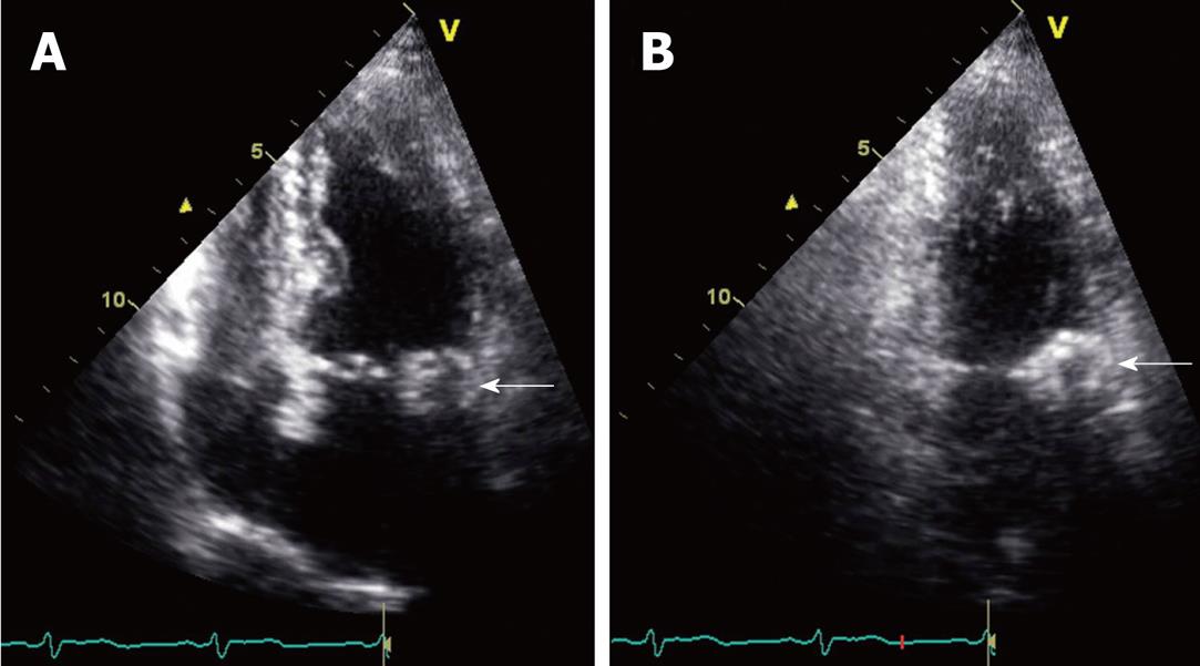
Figure 3 Slightly oblique four-chamber (A) and two-chamber (B) still frame images are obtained from an echocardiogram.
These views demonstrate a mass along the mitral annulus (white arrows). The caseous mitral annular calcifications are seen as an ovoid mass. A difference between the relatively echolucent center and the more echogenic periphery of this structure is perceptible. This is consistent with the centrally liquefied calcium and the peripheral more dense calcium observed on other modalities.
- Citation: Shriki J, Rongey C, Ghosh B, Daneshvar S, Colletti PM, Farvid A, Wilcox A. Caseous mitral annular calcifications: Multimodality imaging characteristics. World J Radiol 2010; 2(4): 143-147
- URL: https://www.wjgnet.com/1949-8470/full/v2/i4/143.htm
- DOI: https://dx.doi.org/10.4329/wjr.v2.i4.143


