Copyright
©2010 Baishideng Publishing Group Co.
World J Radiol. Apr 28, 2010; 2(4): 143-147
Published online Apr 28, 2010. doi: 10.4329/wjr.v2.i4.143
Published online Apr 28, 2010. doi: 10.4329/wjr.v2.i4.143
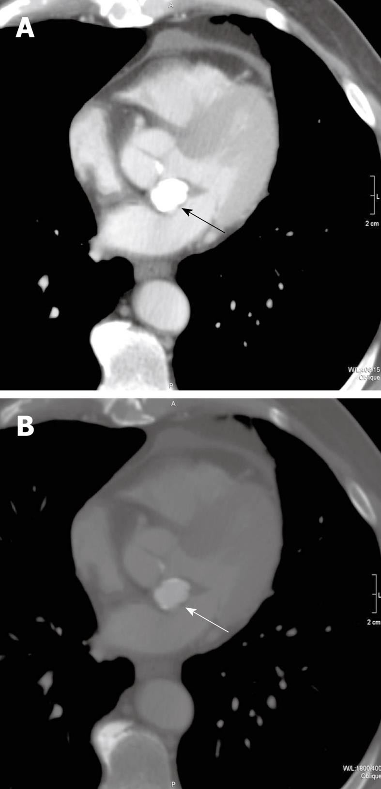
Figure 1 Transverse, non-gated, post-contrast CT images at a level through the heart with mediastinal (A) and bone (B) windows and level settings shown (kVp = 120, mAs = 214, DFOV = 314 mm).
Caseous mitral annular calcifications are noted in the aorto-atrial septum (arrows). On the mediastinal window and level settings, the mass shows homogeneous hyperattenuation which cannot be differentiated from other calcific structures. When the window and level settings are adjusted, there is a rim of peripheral calcification with central, homogeneous hyperattenuation. This mass was stable in comparison to CT scans before and after this study (not shown).
- Citation: Shriki J, Rongey C, Ghosh B, Daneshvar S, Colletti PM, Farvid A, Wilcox A. Caseous mitral annular calcifications: Multimodality imaging characteristics. World J Radiol 2010; 2(4): 143-147
- URL: https://www.wjgnet.com/1949-8470/full/v2/i4/143.htm
- DOI: https://dx.doi.org/10.4329/wjr.v2.i4.143


