Copyright
©2010 Baishideng.
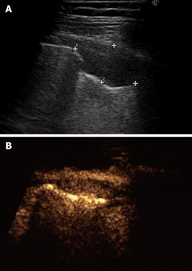
Figure 15 Pleural metastasis.
A: B-mode sonogram shows a hypoechoic lesion resembling either a solid mass or a homogeneously echoic saccate effusion; B: Contrast-enhanced sonography shows contrast enhancement of the lesion 20 s after bolus injection of ultrasound contrast agent. Sonographically-guided biopsy confirmed metastasis from breast carcinoma.
- Citation: Sartori S, Tombesi P. Emerging roles for transthoracic ultrasonography in pleuropulmonary pathology. World J Radiol 2010; 2(2): 83-90
- URL: https://www.wjgnet.com/1949-8470/full/v2/i2/83.htm
- DOI: https://dx.doi.org/10.4329/wjr.v2.i2.83


