Copyright
©2010 Baishideng.
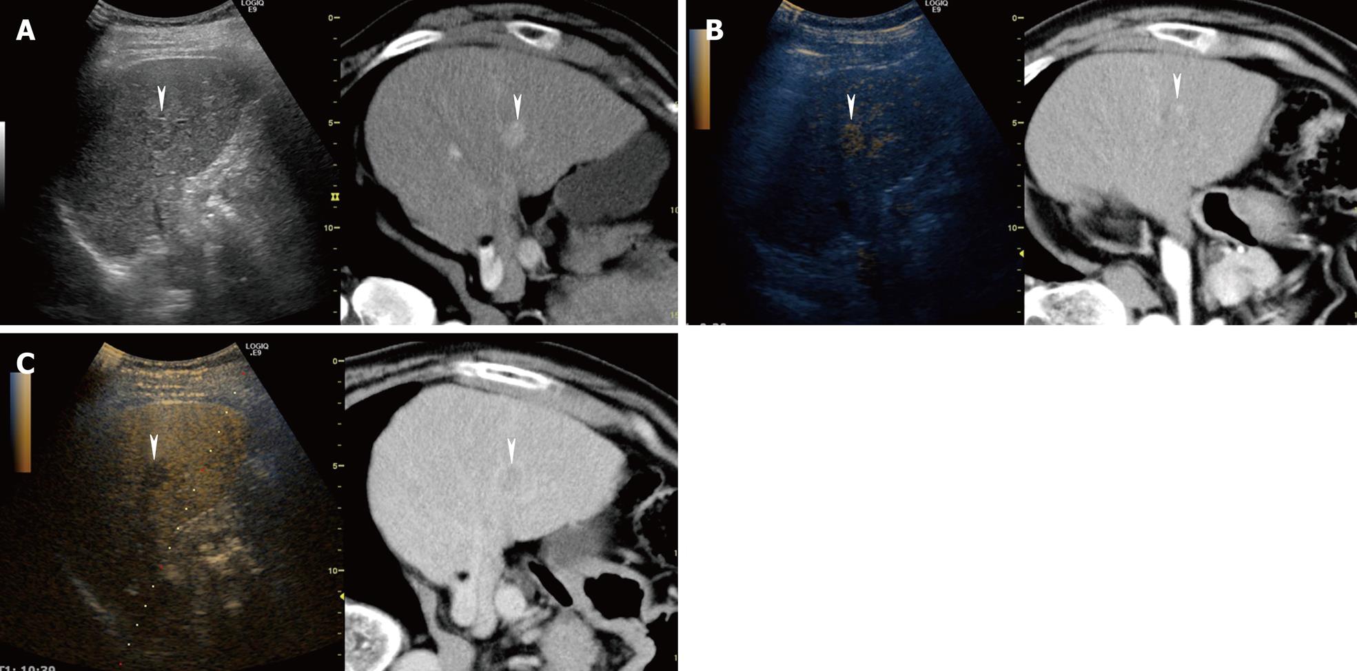
Figure 8 A 66-year-old man with newly developed HCC (maximum diameter 15 mm) in segment V.
A: Fusion image combining arterial phase contrast-enhanced CT (right side) and conventional US (left side). Due to advanced liver cirrhosis, the echogenicity of the liver parenchyma is heterogeneous. Arterial phase contrast-enhanced CT shows a high attenuation area in segment V (arrowheads). Arterial phase contrast-enhanced CT as the reference standard, allows conventional US to detect the target HCC lesion easily; B: Fusion image combining arterial phase contrast-enhanced CT (right side) and early phase Sonazoid-enhanced US at a low MI (left side). Early phase Sonazoid-enhanced US at a low MI shows a homogeneous enhancement in segment V (arrowhead). This enhanced area corresponds well to a high attenuation area, as shown on the arterial phase contrast-enhanced CT image (arrowhead); C: Fusion image combining late phase contrast-enhanced CT (right side) and late phase Sonazoid-enhanced US at a low MI (left side). Late phase Sonazoid-enhanced US at a low MI shows a perfusion defect in a viable HCC lesion (arrowhead). This perfusion defect corresponds well to a low attenuation area, as shown on the late phase contrast-enhanced CT.
- Citation: Numata K, Luo W, Morimoto M, Kondo M, Kunishi Y, Sasaki T, Nozaki A, Tanaka K. Contrast enhanced ultrasound of hepatocellular carcinoma. World J Radiol 2010; 2(2): 68-82
- URL: https://www.wjgnet.com/1949-8470/full/v2/i2/68.htm
- DOI: https://dx.doi.org/10.4329/wjr.v2.i2.68


