Copyright
©2010 Baishideng.
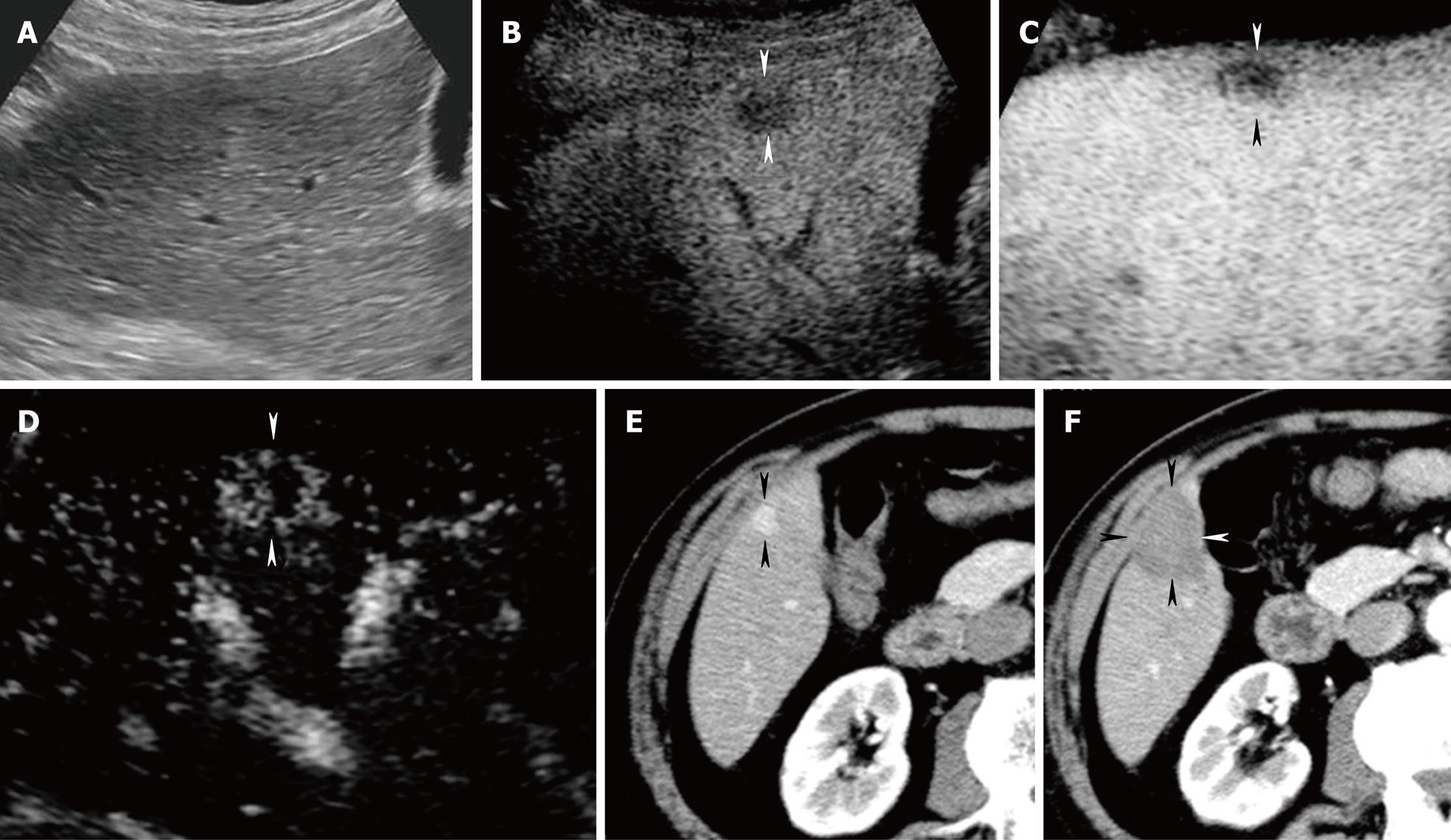
Figure 5 A 67-year-old woman with newly developed HCC (maximum diameter 12 mm) in segment V.
A: Conventional US shows no evidence of tumor; B: Late phase Sonazoid-enhanced US by CPI mode at a low MI shows a perfusion defect (arrowheads); C-D:Late phase Sonazoid-enhanced US by CHA mode on a high MI intermittent image shows a perfusion defect (C) (arrowheads). Intratumoral vessels (D) are seen later (arrowheads). This lesion was treated with percutaneous RFA guided by late phase Sonazoid-enhanced US by CPI mode at a low MI; E-F: Arterial phase contrast-enhanced CT shows a high attenuation area in segment V before RFA (arrowheads) (E). Arterial phase contrast-enhanced CT obtained 4 weeks after treatment shows a low attenuation area (arrowhead) (F). This low attenuation area is larger than that of high attenuation seen in E.
- Citation: Numata K, Luo W, Morimoto M, Kondo M, Kunishi Y, Sasaki T, Nozaki A, Tanaka K. Contrast enhanced ultrasound of hepatocellular carcinoma. World J Radiol 2010; 2(2): 68-82
- URL: https://www.wjgnet.com/1949-8470/full/v2/i2/68.htm
- DOI: https://dx.doi.org/10.4329/wjr.v2.i2.68


