Copyright
©2010 Baishideng.
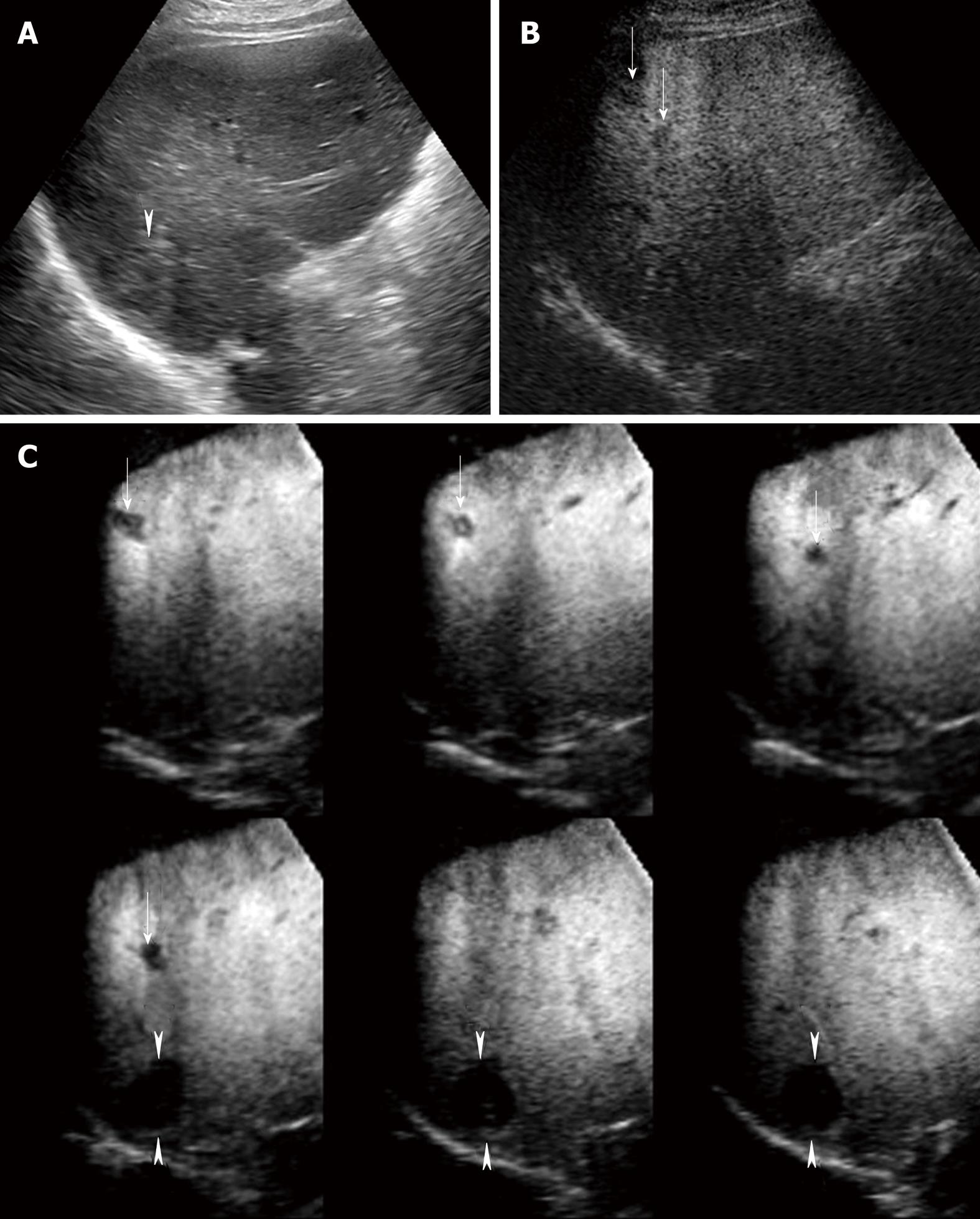
Figure 3 A 75-year-old man with multiple HCC lesions (maximum diameter 22 mm, 12 mm and 10 mm, respectively) in segment VI.
A: Conventional US shows one hyper-echoic HCC lesion alone; B: Late phase Sonazoid-enhanced US by CPI mode at a low MI shows two perfusion defects not detected by conventional US (arrows). However, one hyper-echoic lesion located in the deep portion far from the skin surface cannot be visualized by Sonazoid-enhanced US by CPI mode at a low MI; C: Late phase Sonazoid-enhanced 3D US by CHA mode at a high MI shows three HCC lesions as clear perfusion defects, as depicted on tomographic ultrasound images in plane A, which can be translated from front to back (arrows and arrowheads). The hyper-echoic HCC lesion which was not detected by Sonazoid-enhanced US at a low MI is clearly seen (arrowheads).
- Citation: Numata K, Luo W, Morimoto M, Kondo M, Kunishi Y, Sasaki T, Nozaki A, Tanaka K. Contrast enhanced ultrasound of hepatocellular carcinoma. World J Radiol 2010; 2(2): 68-82
- URL: https://www.wjgnet.com/1949-8470/full/v2/i2/68.htm
- DOI: https://dx.doi.org/10.4329/wjr.v2.i2.68


