Copyright
©2010 Baishideng Publishing Group Co.
World J Radiol. Dec 28, 2010; 2(12): 468-471
Published online Dec 28, 2010. doi: 10.4329/wjr.v2.i12.468
Published online Dec 28, 2010. doi: 10.4329/wjr.v2.i12.468
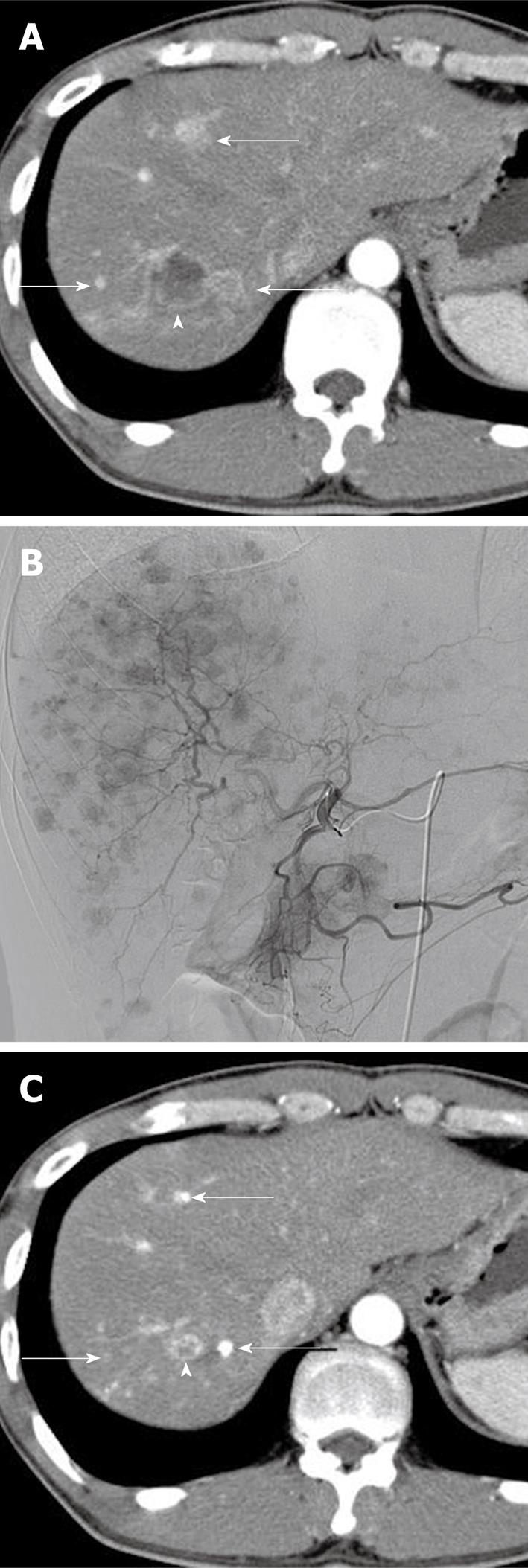
Figure 1 Images obtained in the case of a 38-year-old man with systemic chemotherapy-resistant multiple hepatic metastases from a pancreatic neuroendocrine tumor.
A: Arterial phase image of contrast-enhanced computed tomography (CT) shows well-enhanced metastatic tumors (arrows) as well as a hypodense tumor due to cystic degeneration with marginal enhancement (arrowhead); B: A selective hepatic angiogram delineates innumerable hypervascular metastatic tumors throughout the liver; C: Arterial phase image of contrast-enhanced CT at 3 mo after chemoembolization with miriplatin-iodized oil suspension demonstrates a significant reduction in the size of all lesions, with compact accumulation of iodized oil in the hypervascular tumors (arrows). It should be noted that the tumor with cystic degeneration was also reduced in size (arrowhead).
- Citation: Iwazawa J, Ohue S, Yasumasa K, Mitani T. Transarterial chemoembolization with miriplatin-lipiodol emulsion for neuroendocrine metastases of the liver. World J Radiol 2010; 2(12): 468-471
- URL: https://www.wjgnet.com/1949-8470/full/v2/i12/468.htm
- DOI: https://dx.doi.org/10.4329/wjr.v2.i12.468


