Copyright
©2010 Baishideng Publishing Group Co.
World J Radiol. Oct 28, 2010; 2(10): 405-409
Published online Oct 28, 2010. doi: 10.4329/wjr.v2.i10.405
Published online Oct 28, 2010. doi: 10.4329/wjr.v2.i10.405
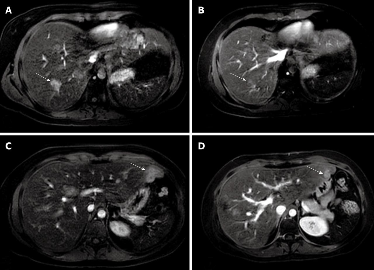
Figure 4 Eovist-enhanced axial magnetic resonance imaging demonstrating enhanced focal nodular hyperplasia lesions in the right liver lobe in arterial phase before treatment (arrow) (A), control magnetic resonance imaging 6 months later showing significantly decreased size of focal nodular hyperplasia lesions (arrow) (B), enhanced focal nodular hyperplasia lesions in the left liver lobe in the arterial phase before treatment (arrow) (C), and significantly decreased size of lesions in the left liver lobe at 6 month follow-up (arrow) (D).
- Citation: Kayhan A, Venu N, Lakadamyalı H, Jensen D, Oto A. Multiple progressive focal nodular hyperplasia lesions of liver in a patient with hemosiderosis. World J Radiol 2010; 2(10): 405-409
- URL: https://www.wjgnet.com/1949-8470/full/v2/i10/405.htm
- DOI: https://dx.doi.org/10.4329/wjr.v2.i10.405


